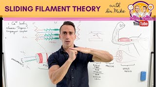
Note that calcium ions do not enter the t-tubule from the SR. Calcium ions diffuse out of the SR through the Ca-release channels into the sarcoplasm where they directly initiate the cross-bridge cycle upon binding to troponin on the thin filament
I’ve managed to struggle less, decrease my time consumption on a single topic, prepare the perfect notes for any upcoming exams, i showed less effort watching your videos than studying alone but still managed to get a better outcome and i feel so grateful towards you.
Technically the t-tubules are dips into the muscle fiber, from the sarcolemma, they don't run around the myofibril. Although you are correct that the SR are around each myofibril. The SR and the t-tubule are in contact through the DHPR receptor on the t-tubule and the RYR receptor on the SR. When the voltage runs down the t-tubule the DHPR response to the voltage by changing 'shape' which in turn cause the RYR to open to allow Calcium to flow into to cytoplasm. Calcium never enters the t-tubule. Calcium binds to troponin which is located on the thin filaments. I should add that your videos are my go to videos that I suggest to my students. Nice artwork!
+UnseenNature Agreed with all of this. Ca2+ never actually enters the t-tubules as they are technically invaginations of the extracellular space. Love the vidoes.
Dude, I have no words to thank you for these videos. Not only you ilustrate the information gathered in books and classes perfectly but you also do it in a very didatic way. You've got an amazing talent, keep it on!
"Sarco" not "sacro", and the transverse tubules are invaginations of the sarcolemma, so the Ca ions do not release into the t-tubules, but the voltage-gated protein receptor between the SR and the t-tubule releases the Ca into the sarcoplasm through the sides of the gate i.e. The gaps in the triad. Apart from that, love the drawing.
I’ve been watching your channel for a while now to help me with my study, I’m a medical student and I’ve been having a hard time making sense out of my professor bad explanation, with you amazing help I managed to pass my exams only by watching your videos and only reading my materials, thank you so so much hope to see more of your videos coming to help all of the medical students out there in need wish you best of luck.🤍🙏🏻
You just saved me. I haven't understood this, until you explained it with your drawing. I just might get a good grade on my final exam. I can't thank you enough. I am going to share this video with my instructor.
Your drawings are BRILLIANT. Makes me understand it much easier. That's the thing about exam boards they teach you about the important things but never go into detail like you did.
Thank you soooooo much for taking the time and effort to put these videos together. It has definitely made this material easier to visualize and understand.
What you call a motor unit is actually a motor end plate. The motor unit is a collection of all end plates innervated by a motor neurone plus the neurone itself.
Its key to note that when the action potential travels down the t-tubules, a receptor ON the t-tubules (DHP receptor) opens what is known as RyR receptors on the Sarcoplasmic reticulum. and from there Calcium ions go STRAIGHT into the cytoplasm and bind to troponin and not necessarily travel down the t-tubules. Everything else however is nicely stated!! nicely done
Omg I was so confused, but you explained this perfectly. What a relief that now I understand this because I am a visual learner and looking at my notes wasn’t doing anything for me
It is physiologically important for excitation-contraction coupling hat the T-tubules are positioned close to the terminal cisternae of the sarcoplasmic reticulum as the triad or diad arrangement allows physical and functional contact by voltage dependent L-type calcium channels. So, an action potential along the sarcolemma causes calcium channels to open in the terminal cisternae/sarcoplasmic reticulum which enables calcium to move from the sarcoplasmic reticulum into cytoplasm and the intracellular calcium concentration to increase.
A very good brief discussion of Skeletal Muscle Anatomical Physiology as well as the propagation of COntraction. It strengthens and helped me recall the things Guyton told me :)
Lovely art work! You've hit upon a common misconception among students. I like to ignore the historical names given to the proteins involved and simply describe their functions. So think of the T-tubules as conductors - they carry the electrical impulse deep into the cell. Their membranes contain voltage sensitive switches which are activated by change in membrane potential. Those switches then open calcium channels in the membranes of the SR, allowing Calcium ions to flow into the sarcoplasm, initiating the cross-bridge cycling. The calcium pump (Calcium ATPase) also found in the membrane of the SR is always on and will eventually catch up, returning calcium levels in the sarcoplasm to resting levels, and allowing relaxation.
The video is incorrect. The T-tubule contains voltage gated calcium(II) channels, that are mechanically linked to calcium(II) channels in the membrane of the sarcoplasmic reticulum. The action potential opens the voltage gated calcium(II) channels of the T-tubule, resulting in a flux of ECM calcium(II)-ions into the muscle fiber. The mechanical link between the voltage gated ion channels of the T-tubule and the calcium(II) channels of the SR, then causes the latter to open with a flux of calcium(II) into the cytosol from the SR. This great increase of calcium(II) then causes contraction by binding to troponin. Hope this he
When the acetylcholine receptors open in the junctional folds of the motor end plate and open the ion channels to allow the positively-charged sodium ions into the muscle fibre, what happens to them/where do they go once the action potential has been propagated along the sarcoplasmic reticulum..?
@@jamesDJRPM to repolarize there are potassium channels that bring potassium out of the cell in order to repolarize the cell and bring back the resting membrane potential then it becomes more negative than the rmp then na-k atpase pumps return the na ICF to the ECF and potassium ECF to ICF again.
I just have to say you have been a great help in understanding this. The visual imagery was something very helpful that I wasn't getting in class, but this made it a lot easier. Thank you!
Hi, some people have already pointed it out, but ill just say it again, the Ca+ leaves the sacroplasmic reticulum straight onto the filaments, the inside of the t-tubule is part of the extracellular space.
This is a wonderful video. Hitting all the required/important points to introduce to muscle contraction. The best articulation of the topic I've seen thus far. Your drawings greatly assist. Great work,keep it up.
Not just a like but my gosh you are amazing. Awesome presentation everybody should know about this. From people who are learning the basics to people who are learning intricate details amazing.
what an amazing video, had a good idea of muscle contraction and the muscular system but this video really puts it all together. From an RT student, thank you!
Armando, I wish you would come and teach at my Uni. Ive taken to just watching through your video's because your've made learning fun again, thank you so much and keep up the exceptional work!
When the ACh is released, it binds to ACh receptors at the neuromuscular junction, opening ligand-gated Na+ channels (not voltage-gated) that causes depolarization causing the AP to spread. This was a small typo, I thought I'd point out.
Great as always except a small nitpicking: (2:40) "sacrolemma" --> "sarcolemma"; (3:05) "sacromere" --> "sarcomere"; (3:30) "sacroplasmic R." --> "sarcoplasmic R." By the way, calcium ions are not getting into T-tubules but rather into sarcoplasm via the vast network of SR itself, I think.
Thank you for making these videos! I thought I would be lost and confused forever and then I watched this. I may actually pass A&P with a decent grade. Thanks a million!
Real ease of NTs require Ca entry at presynpatic junction. Na entry trough nACHR doesn’t generate action potentials itself ,it cause depolarisation,if it’s above the threshold then it will open Voltage gated Na channels. So first Na entered through inotropic channel i.e nACHR then through Voltage gated channels. T tubules are connected to DHPR Ca channel, acting sites of Amlodepine, which is just adjacent to RyR ( site of Dantrolene - for fatal Malignant hyperthermia secondary to few anaesthetics). Good job anyway.
Thanks for that! I realized that but didn't explain it as well as I was thinking it. Anyways, I really enjoy and appreciate your videos and they really helped me gain a better understanding! Keep up the good work man!
Dude this video was amazing!! Your art was great, made it easier to understand what you were talking about. I thought your flow of the material and the movement of the drawings to your information was great! Thank you for this great study video
I love your videos. Your drawings are great and I'm recommending them to my students in A&P to help them study. HOWEVER, the Calcium does not flow into the T-tubules from the SR. It goes into the sarcoplasm. The T-tubules are projections of the sarcolemma and if the Calcium went into them, it would be going out of the cell.
Not to be a grammar nazi, but i noticed that for "Sarco" IE Sarcoplasm, Sarcolemma, etc You put "Sacro" 'c' before the 'r' which was throwing me off at first because i thought i had been reading it wrong all this time in my text book! Just thought I would point that out! otherwise love the vid it has helped me understand this somewhat confusing and complex subject!


![Oasis - Oasis Live '25 [Official Trailer]](http://i.ytimg.com/vi/gol_JnuEtKM/mqdefault.jpg)


Note that calcium ions do not enter the t-tubule from the SR.
Calcium ions diffuse out of the SR through the Ca-release channels into the sarcoplasm where they directly initiate the cross-bridge cycle upon binding to troponin on the thin filament
I’ve managed to struggle less, decrease my time consumption on a single topic, prepare the perfect notes for any upcoming exams, i showed less effort watching your videos than studying alone but still managed to get a better outcome and i feel so grateful towards you.
Achaaaa g
Technically the t-tubules are dips into the muscle fiber, from the sarcolemma, they don't run around the myofibril. Although you are correct that the SR are around each myofibril. The SR and the t-tubule are in contact through the DHPR receptor on the t-tubule and the RYR receptor on the SR. When the voltage runs down the t-tubule the DHPR response to the voltage by changing 'shape' which in turn cause the RYR to open to allow Calcium to flow into to cytoplasm. Calcium never enters the t-tubule. Calcium binds to troponin which is located on the thin filaments.
I should add that your videos are my go to videos that I suggest to my students. Nice artwork!
+UnseenNature
Agreed with all of this. Ca2+ never actually enters the t-tubules as they are technically invaginations of the extracellular space.
Love the vidoes.
THANK YOU! I was panicking for a sec because the video wasn't the same as what I learned in class.
you are right
sarcoplasm not cytoplasm?
@@abhayxp8062 yes, because sarcos in greek means meat, thats why they use it for the muscle tissue
It's amazing to think that all this happens EVERYTIME you use a skeletal muscle. It's almost overwhelming to wrap your mind around.
Sorry, I didnt really talk about it in this video.
You can watch it here:
Myology - Skeletal Muscle (Sarcomere, Myosin and Actin)
Is this the video for sliding filament theory?
after struggling and leaning muscle contraction for the whole day, I finally got a clear head after your demonstration!! THANK YOU!!
Dude, I have no words to thank you for these videos. Not only you ilustrate the information gathered in books and classes perfectly but you also do it in a very didatic way. You've got an amazing talent, keep it on!
Totally agree with you👌🏼✨
"Sarco" not "sacro", and the transverse tubules are invaginations of the sarcolemma, so the Ca ions do not release into the t-tubules, but the voltage-gated protein receptor between the SR and the t-tubule releases the Ca into the sarcoplasm through the sides of the gate i.e. The gaps in the triad. Apart from that, love the drawing.
yeah was thinkin the same thing
I also noticed he said the word right but then wrote it wrong. Then I had to look it up.
Can you just take all my notes for me? This is beautiful and it makes more sense than anything my professor was trying to say...
Hi beautiful. Can i get your whatsapp number# 😎☺
@Muhammad Baqir I have free notes I can send on email. 56 pages.
@@khalidmehsud54 Har jaga badnaam karte ho tum log. Besharam
Send me !!
@@themetalroofcompany3664 send me!!!
I’ve been watching your channel for a while now to help me with my study, I’m a medical student and I’ve been having a hard time making sense out of my professor bad explanation, with you amazing help I managed to pass my exams only by watching your videos and only reading my materials, thank you so so much hope to see more of your videos coming to help all of the medical students out there in need wish you best of luck.🤍🙏🏻
You just saved me. I haven't understood this, until you explained it with your drawing. I just might get a good grade on my final exam. I can't thank you enough. I am going to share this video with my instructor.
Your drawings are BRILLIANT. Makes me understand it much easier. That's the thing about exam boards they teach you about the important things but never go into detail like you did.
Great Videos!
What We could not understand from hours long lecture you have explained in short time.
Ikdt
Ikr that's what I'm supposed to wrire
Thankyou Armando. Since I found you on youtube, it was easy to pass my exams at medical school.
Thank you soooooo much for taking the time and effort to put these videos together. It has definitely made this material easier to visualize and understand.
Youre pure LEGEND...from Missouri/California....THANKS
What you call a motor unit is actually a motor end plate. The motor unit is a collection of all end plates innervated by a motor neurone plus the neurone itself.
This was amazing! you explained this so well! I did not understand in an hour and thirty minutes in lecture! i understood it in 7 minutes!! thanks
Its key to note that when the action potential travels down the t-tubules, a receptor ON the t-tubules (DHP receptor) opens what is known as RyR receptors on the Sarcoplasmic reticulum. and from there Calcium ions go STRAIGHT into the cytoplasm and bind to troponin and not necessarily travel down the t-tubules. Everything else however is nicely stated!! nicely done
This was explained so well!! Thank you! :) I sometimes got distracted by how incredible the artwork was lol
Very informative and beautifully illustrated. Thank you for posting!
Great drawings. I can tell a lot of work was put into this.
7:05 CORRECTION : Calcium ions are released from SR to sarcoplasm and bind to troponin C because actin and myosin filaments are in the sarcoplasm.
You've created a very practical and enjoyable way to learn biology. Very Well Done!
This series of medical videos is an excellent way of conveying information. Great and innovative effort. Keep up the good work.
Omg I was so confused, but you explained this perfectly. What a relief that now I understand this because I am a visual learner and looking at my notes wasn’t doing anything for me
This was the amazing. I don't often see videos that are are so easy to follow and simplify the complicated process as well as you did. Thank you.
WHEW! art and illustration saves the day yet again. sometimes artists only learn from other artists. nice work.
Great illustrations, and it makes it so much Easier to understand. thank you Armando
It is physiologically important for excitation-contraction coupling hat the T-tubules are positioned close to the terminal cisternae of the sarcoplasmic reticulum as the triad or diad arrangement allows physical and functional contact by voltage dependent L-type calcium channels. So, an action potential along the sarcolemma causes calcium channels to open in the terminal cisternae/sarcoplasmic reticulum which enables calcium to move from the sarcoplasmic reticulum into cytoplasm and the intracellular calcium concentration to increase.
A huge THANKS! One of the clearest and easy to follow descriptions I've seen. Really helping my studies...
Thank-you for taking the time to make this, it has just made my life a lot easier
oh.. life?
⊙.☉
I am in love with your teaching skills and artworks
A very good brief discussion of Skeletal Muscle Anatomical Physiology as well as the propagation of COntraction. It strengthens and helped me recall the things Guyton told me :)
Great video...
T-tubule propagates action potential towards sarcoplasmic reticulum (which contains Ca2+) and causes to Ca ion channels on SR to open.
Your explanation and drawings were very helpful! Thank you!
Sir you are the greatest teacher ever
Lovely art work! You've hit upon a common misconception among students. I like to ignore the historical names given to the proteins involved and simply describe their functions. So think of the T-tubules as conductors - they carry the electrical impulse deep into the cell. Their membranes contain voltage sensitive switches which are activated by change in membrane potential. Those switches then open calcium channels in the membranes of the SR, allowing Calcium ions to flow into the sarcoplasm, initiating the cross-bridge cycling. The calcium pump (Calcium ATPase) also found in the membrane of the SR is always on and will eventually catch up, returning calcium levels in the sarcoplasm to resting levels, and allowing relaxation.
Perfect details after some prior revision, it maybe daunting to watch these videos with no studying. Well done
In 7 minutes i learned more than reading the book for an hour. Thank you.
the Ca++ doesnt get out from the SR to de T-Tubule and then to de citoplasm; it gets out from the SR directly to de citoplasm (sarcoplasm)
I really like what you did here! Very clean and easy explanation.
One minor faulty though: Sarcomere, not sacromere..
Keep up the good work!
Sir I hv studied dis chaptr frm one of d best faculty of india...and found u d best..awsm..kindly upload more pls....:)
The video is incorrect. The T-tubule contains voltage gated calcium(II) channels, that are mechanically linked to calcium(II) channels in the membrane of the sarcoplasmic reticulum. The action potential opens the voltage gated calcium(II) channels of the T-tubule, resulting in a flux of ECM calcium(II)-ions into the muscle fiber. The mechanical link between the voltage gated ion channels of the T-tubule and the calcium(II) channels of the SR, then causes the latter to open with a flux of calcium(II) into the cytosol from the SR. This great increase of calcium(II) then causes contraction by binding to troponin. Hope this he
When the acetylcholine receptors open in the junctional folds of the motor end plate and open the ion channels to allow the positively-charged sodium ions into the muscle fibre, what happens to them/where do they go once the action potential has been propagated along the sarcoplasmic reticulum..?
@@jamesDJRPM to repolarize there are potassium channels that bring potassium out of the cell in order to repolarize the cell and bring back the resting membrane potential then it becomes more negative than the rmp then na-k atpase pumps return the na ICF to the ECF and potassium ECF to ICF again.
Voltage gated calcium channel L type the first group of VGCachannel* in cardiac muscle and skeletal muscle.
What is the name of the acetylcholine receptor? he didn't mention it!
I just have to say you have been a great help in understanding this. The visual imagery was something very helpful that I wasn't getting in class, but this made it a lot easier. Thank you!
BEST explanation ever. Love the art and love the civilized British accent
its amazing how a simple muscle can be more complex than a circuit board
Hi, some people have already pointed it out, but ill just say it again, the Ca+ leaves the sacroplasmic reticulum straight onto the filaments, the inside of the t-tubule is part of the extracellular space.
I didn't understand any of this in my 4h physiology class and you just explained it all in 7:40 minutos
I wouldn't graduate if you didnt make such beautiful work! Excellent videos!
This is a wonderful video. Hitting all the required/important points to introduce to muscle contraction. The best articulation of the topic I've seen thus far. Your drawings greatly assist. Great work,keep it up.
This is explained so clearly unlike a few of my lectures, its great! Thanks for posting, i'll be watching more soon!
thank you so much i have my semester tomorrow really helpful. always last minute studies will help a lot.
beautifully explained...
my teacher explained this for 2 hrs...
This video is perfectly explained. Thank you
You just made ma physiology class much easier. tnx a lot. We gonna expect more videos from you. Thank you again.
Thank you for posting these videos! Really helping with my revision for my biology exams !
Not just a like but my gosh you are amazing. Awesome presentation everybody should know about this. From people who are learning the basics to people who are learning intricate details amazing.
what an amazing video, had a good idea of muscle contraction and the muscular system but this video really puts it all together. From an RT student, thank you!
Sir you have a gift, I cant thank you enough!
Excellent. Concise and easy to follow.
I never leave comments but just want to thank you so much! Great teaching and terrific drawings. I agree the notes would be useful as a download.
ok, your drawing skill is insane, congratulation!
Really appreciate it! Good work Armando.
Can´t thank you enough. Every one of your videos are great, please keep up your amazing work!
Great video. It helped me a lot during my A and P I. Thanks!
Your artwork and explanation was extraordinary. Thank you so, so much!
I like your hand writing and your draw,Nice job.
Armando, I wish you would come and teach at my Uni. Ive taken to just watching through your video's because your've made learning fun again, thank you so much and keep up the exceptional work!
Excellent video. Concept Very well explained with good drawings / diagrams. Much much easier than reading & understanding directly from the text. 👍
Heads off such a great teacher you're legend sir!!!
HONESTLY 💯
hey Armando
nyc drawing and u explain the things in the easy way
very very helpful! best combination of animation and explanation ive seen yet. thank you
Incredible! Your videos piece all of this information together so well. Thank you so much for putting this out here!
When the ACh is released, it binds to ACh receptors at the neuromuscular junction, opening ligand-gated Na+ channels (not voltage-gated) that causes depolarization causing the AP to spread. This was a small typo, I thought I'd point out.
Great as always except a small nitpicking: (2:40) "sacrolemma" --> "sarcolemma"; (3:05) "sacromere" --> "sarcomere"; (3:30) "sacroplasmic R." --> "sarcoplasmic R." By the way, calcium ions are not getting into T-tubules but rather into sarcoplasm via the vast network of SR itself, I think.
so nice of u Armando....may you live long dear. you and ninja nerd... what an effort yaaarrr....👍👍👍
Killing the lesson man. This is spot on to my last human physio lecture. Keep up the good work.
Thank you for making these videos! I thought I would be lost and confused forever and then I watched this. I may actually pass A&P with a decent grade. Thanks a million!
Great job! This is my favorite part of A&P!
Amazing explaination with amazing diagrams. Creating a lot of interest in studing. Thank u soooooo...... much. We love u a lot. 😍😘😘😘
This happened me so much understand the concept of muscle contractions!! life saver!!!
Real ease of NTs require Ca entry at presynpatic junction.
Na entry trough nACHR doesn’t generate action potentials itself ,it cause depolarisation,if it’s above the threshold then it will open Voltage gated Na channels. So first Na entered through inotropic channel i.e nACHR then through Voltage gated channels.
T tubules are connected to DHPR Ca channel, acting sites of Amlodepine, which is just adjacent to RyR ( site of Dantrolene - for fatal Malignant hyperthermia secondary to few anaesthetics).
Good job anyway.
Thanks for that! I realized that but didn't explain it as well as I was thinking it. Anyways, I really enjoy and appreciate your videos and they really helped me gain a better understanding! Keep up the good work man!
Hey Armando, great videos man! Very nicely done.
Dude this video was amazing!! Your art was great, made it easier to understand what you were talking about. I thought your flow of the material and the movement of the drawings to your information was great! Thank you for this great study video
Beautifully explained ...... You are amazing sire 😃
I love your videos. Your drawings are great and I'm recommending them to my students in A&P to help them study. HOWEVER, the Calcium does not flow into the T-tubules from the SR. It goes into the sarcoplasm. The T-tubules are projections of the sarcolemma and if the Calcium went into them, it would be going out of the cell.
Helped me greatly during exam. Thank you.
Excellent diagramatic representation 👏
Great video, really helpful and easy to understand
Very informative in easiest way..... Respect from Pakistan
wow you explained way better in 7 min then what my biology teacher took 3 days to explain
Yes Mawi C. Watch the Armando's Neuromuscular Junction episode for a more thorough explanation of that process.
Thank you, you've helped my physiology class
awesome awesome awesome.... best way to teach .... concept got cleared by dis demonstration 😍... thnx aloooooot
your the best. THANK YOU for making this so easy to understand !!!
You make it so simple , Thank you very much
Isn't Calcium what triggers ACh to exit the axon terminal via exocytosis?
Hi
Excellent work for better understanding..!!
Not to be a grammar nazi, but i noticed that for "Sarco" IE Sarcoplasm, Sarcolemma, etc You put "Sacro" 'c' before the 'r' which was throwing me off at first because i thought i had been reading it wrong all this time in my text book! Just thought I would point that out!
otherwise love the vid it has helped me understand this somewhat confusing and complex subject!
Yes..