Tissue Sectionning using an ultra- microtome

HTML-код
- Опубликовано: 12 сен 2024
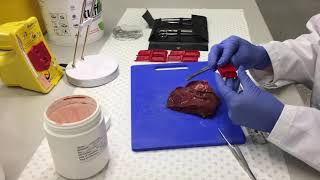
- This video shows how a resin block containing a sample is cut and processed using an ultra-microtome. First semi-thin (400nm) are cut with a glass knife and stained blue with Toluidine Blue to check that we are in the right place. Then thin sections (70nm) are cut and collected on a grid. These sections are then stained for transmission electron microscopy (TEM) as shown in a different video.


Excellent video.
That model of ultramicrotome is beautiful. I think it is a Reichert Jung OMU3. The macro images showing slices being coaxed onto a grid are very nice.
❤ nice