Personally speaking it's the best flow cytometry introductory video so far. The mechanisms are explained in a clear yet informative way. Details like channel resolution, flow structure etc are included which is scarce in other similar videos. Thanks!
Watched a lot of videos before coming across yours, and now i can finally sat I understand how it works. Thank you keep making great content like this.
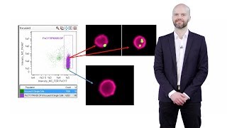
Many thanks for great explanation but Can I have a question? What does the colors of the dots indicate (as in 28:55). Is it the density? Like the number of cells in red area will higher than yellow one, and yellow > green > blue right? sorry for my poor english
Can we apply two different lazers (e.g., FITC and PE) at the same time to excite cells, and then detect and sort only cells that are exicted by both lazers, and get data from computer? This would be the same effect by Image J. (e.g. Getting FITC and PE images, respectively from fluorescence microscope and then merge them, which generates yellow pseudo color)
PE is a fluorophore that fluoresces at ~580 nm FITC is a fluorophore that fluoresces at ~500 nm CD4 is a helper T cell marker. Functionally, it is the TCR coreceptor for helper T cells CD3 is a T cell marker. Functionally, it makes up the signalling part of TCR complexes Hyphens indicate covalent linkage. I guess it would be more correct to say PE-αCD4 and FITC-αCD3 to show that these are antibodies for these cell markers.
A question, In the Hydrodynamic focusing, Is the sheath fluid merging with the blood sample (which already diluted) if yes, then the dilution ratio will be changed? and if no, How they are not merging ! TIA
I am not completely sure but I think they don't cuz the idea of hydrodynamic focusing is to kind of create a channel based on the sheath fluid. This is what I found in Wikipedia: "The sample is injected into the middle of the sheath flow. If the two fluids differ enough in their velocity or density, they do not mix: they form a two-layer stable flow."
www.google.com/url?sa=t&source=web&rct=j&url=expertcytometry.com/fluidics/&ved=2ahUKEwju44b8x_7jAhUto1kKHRzWCEgQFjAQegQICRAB&usg=AOvVaw1ePLa1Lp5dRhmX4D-3Pzi9 ok no they don't mix, confirmed hahaha


Personally speaking it's the best flow cytometry introductory video so far. The mechanisms are explained in a clear yet informative way. Details like channel resolution, flow structure etc are included which is scarce in other similar videos. Thanks!
Thank you iBiology and Dr. Paulsen for this very useful and information-rich video!
Watched a lot of videos before coming across yours, and now i can finally sat I understand how it works. Thank you keep making great content like this.
I watched this when I was learning flow and continue to send it to other grad students. Such a great video!
Excellent video. I wish the one was released in 2010 when I started my PhD. Had I seen this one, I would had not made so many avoidable mistakes.
Very well covered! Great to learn a bit more of the fundamentals of it beyond just going through the motions of using it
Dang! That was fascinating and scarily understandable, thank you!!!
it is very informative and simpified. many thanks malte
Thank you for the great explaination!
Really nice and clear explanation. Congratulation. Looking forward for a video into multiparametric analysis of data
I loved this video, it's very nicely explained and the animations are great. Just one minor comment that 2^4 goes to 16 channels. Thanks for sharing 💕
Excellent video
Very nice video! Thanks!
The cell orientation doesn't affect the scattering? Or are the cells oriented in somehow? Maybe I missed something.
thank you, this was very helpful
SO nice and helpful
Great information and video
Many thanks for great explanation but
Can I have a question?
What does the colors of the dots indicate (as in 28:55). Is it the density? Like the number of cells in red area will higher than yellow one, and yellow > green > blue right?
sorry for my poor english
awesome mate. Thanks
Awesome video! Subtitle required. I can't find Auto-subtitle option.
great video
Can we apply two different lazers (e.g., FITC and PE) at the same time to excite cells, and then detect and sort only cells that are exicted by both lazers, and get data from computer?
This would be the same effect by Image J. (e.g. Getting FITC and PE images, respectively from fluorescence microscope and then merge them, which generates yellow pseudo color)
well done!
Amazing
GREAT!
Thank you
Thanks Sir you and your video is very nice. Your explanation method is best. Sir please explain between PE-CD4 and FITC-CD3 .
PE is a fluorophore that fluoresces at ~580 nm
FITC is a fluorophore that fluoresces at ~500 nm
CD4 is a helper T cell marker. Functionally, it is the TCR coreceptor for helper T cells
CD3 is a T cell marker. Functionally, it makes up the signalling part of TCR complexes
Hyphens indicate covalent linkage. I guess it would be more correct to say PE-αCD4 and FITC-αCD3 to show that these are antibodies for these cell markers.
Super informative video! Just wondering but isn't it 45 bivariate combinations with 10 antibodies?
Awesome vedieo
A question, In the Hydrodynamic focusing, Is the sheath fluid merging with the blood sample (which already diluted) if yes, then the dilution ratio will be changed? and if no, How they are not merging ! TIA
I am not completely sure but I think they don't cuz the idea of hydrodynamic focusing is to kind of create a channel based on the sheath fluid. This is what I found in Wikipedia: "The sample is injected into the middle of the sheath flow. If the two fluids differ enough in their velocity or density, they do not mix: they form a two-layer stable flow."
www.google.com/url?sa=t&source=web&rct=j&url=expertcytometry.com/fluidics/&ved=2ahUKEwju44b8x_7jAhUto1kKHRzWCEgQFjAQegQICRAB&usg=AOvVaw1ePLa1Lp5dRhmX4D-3Pzi9 ok no they don't mix, confirmed hahaha
dilution/not-dilution makes no difference. The cells will be assessed as they go through the laser and that is the info you need.
one size FITC all
Mn wojoohahom ta3refoonahom. Even their dialogue.. No need for cytometry