EXTRAVASATION

HTML-код
- Опубликовано: 10 июл 2024
- / neuralacademy
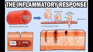
The inflammatory response is initiated within hours of infection or wounding and is triggered by physical damage to tissues or the presence of pathogens. During inflammation, there is cytokine release, vasodilation, and recruitment of leukocytes. Cytokines are substances secreted by cells of the immune system that affect other cells. Chemokines are a type of cytokine that induce directed movement of cells. Vasodilation is the dilation of blood vessels. So how do the leukocytes enter inflamed tissues?
Usually, leukocytes travel in the center of blood vessels, where blood flows the fastest. The first step of leukocyte recruitment into infected tissues is dilation of blood vessels, resulting in slower blood flow. This allows leukocytes to interact with the vascular endothelium. Now, leukocytes need to stick to the blood vessel walls.
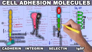
During inflammation, cytokines cause changes in the adhesion molecules on the endothelial cells, as well as the adhesion molecules expressed by leukocytes. Three kinds of adhesion molecules are important for leukocyte recruitment: Selectins, Intracellular Adhesion Molecules (or ICAMs), and Leukocyte Integrins.
Selectins are membrane glycoproteins that bind specific carbohydrate groups. They are expressed on activated endothelium and initiate endothelium-leukocyte interactions by binding to fucosylated oligosaccharide ligands on passing leukocytes. ICAMs are single-pass membrane proteins that allow for tighter adhesion of the leukocytes to the endothelium.
Leukocyte integrins are composed of two transmembrane protein chains, α and β, of which there are numerous types. Subsets of integrins have a common β chain partnered with different α chains. Leukocyte integrins important for extravasation are αLβ2 and αMβ2, which can bind to both ICAM-1 and ICAM-2. Integrins also allow for the convenient distinguishing of different cell types. Dendritic cells, macrophages, and monocytes feature different integrin α chains and thus display distinct β2 integrins on their surface.
Migration of leukocytes out of blood vessels - extravasation. Extravasation has four steps: Rolling Adhesion, Tight Binding, Diapedesis, and Migration.
1. ROLLING ADHESION The initially weak adhesion between leukocytes and the vascular endothelium involves selectins (P-selectin and E-selectin). P-selectin appears on the endothelial cell surface within minutes of exposure to histamine, which is released by mast cells, or exposure of the endothelium to TNF-α or LPS.
TNF-α is a cytokine produced by macrophages upon pathogen detection and causes endothelial activation. Activated endothelial cells rapidly externalize granules called Weibel-Palade bodies, which contain preformed P-selectin. TNF-α and LPS also induce the synthesis of a second selectin, E-selectin, which appears on the endothelial cell surface a few hours later.
These selectins recognize the sulfated sialyl-LewisX moiety of certain leukocyte glycoproteins. P-selectin and E-selectin interact with these glycoproteins, allowing the leukocytes to adhere reversibly to the vessel wall, so they can “roll” along the endothelium. Without this initial weak adhesion, the stronger adhesion in the next step in extravasation can’t happen.
2. TIGHT BINDING Tight binding relies on interactions between leukocyte integrins and adhesion molecules on the endothelium, such as ICAM-1 and ICAM-2. Leukocyte integrins normally bind their ligands only weakly, but chemokines bound to proteoglycans on the surface of endothelial cells bind to specific chemokine receptors on the leukocyte and signal the cell to trigger a conformational change in the integrins on the rolling leukocyte, greatly increasing the adhesive abilities of the leukocyte. As a result, the leukocyte can attach firmly to the endothelium and the rolling stops.
3. DIAPEDESIS In this step, the leukocyte extravasates, or crosses, the endothelial wall. This again involves the leukocyte integrins, as well as further adhesive interactions involving an immunoglobulin-related molecule called PECAM or CD31, expressed both on the leukocyte and at the intercellular junctions of endothelial cells. Next, the leukocyte penetrates the basement membrane with the aid of enzymes that break down extracellular matrix proteins. The movement through the basement membrane is known as diapedesis, and once the leukocyte has crossed it, it is now in the subendothelial tissues.
4. MIGRATION Migration of leukocytes through tissues occurs under the influence of chemokines produced at the site of infection. A concentration gradient of chemokines is formed along which the leukocyte can migrate to the focus of infection.
To end off, note that, even in uninfected regions of the body, circulating monocytes are continuously leaving the blood and entering tissues, where they become resident macrophages. They do so when they adhere to ICAM-2, which is expressed at low levels by unactivated endothelium.


I'm probably late to this comment's section and I don't comment on RUclips videos very often. I've been trying to solve this problem on Uworld questionnaire bank and grasp the extravasation concept for almost about an hour now. Tired and defeated, I just decided to get out of my reading comfort zone and watch a video instead. Took me only 5 minutes (the length of this video) to understand it. I can't thank this channel enough for the upload; the entire thing had really started to get on my nerves. You guys are life savers.
I have been struggling with Histology all semester and our blood section was entirely self-taught. We were only given a single powerpoint slide explaining this complex process and I felt hopeless. This video made it all click and make sense, thank you so much!
I'm about to have my immunology exam for my pharmaceutical degree in about a month and your videos really help me.
Great Job 👍
i spent hours to understand this operation before watching your video its really amazing !
glad you came back at the end
i was searching for you
now i'm complete
thanks @neural academy
Just damn well explained bro
This video is SO good. So illustrative
Have my Biomedical sciences immunology exam in a week and this really helped :)
Thank u for the animated explanation it helps me to understang robbins in general pathology. I am a incoming 2nd year medical student. . . Thank u so much
This was crazy helpful! Thank you :D
I love it, bro.Thank you so much.
Perfect explanation
What an incredibly awesome video !
Applauses from Korea THANKS
Good video, thank you. I instantly subscribed.
Damn, best explanation so far! Thank you
quick and to the point.
Thanks for explaining so well !
It's perfect, good concepts in short and simple words.
I'm gonna cry .. This is perfect !!!!!!!!!
Soooooo truuuuu he is amazing
Thank you so so much for the explanation
Love this, thank you so much
My daughter was discouraged, having to learn anatomy on her own from a textbook. This video put a smile on her face and I am sure she will watch it again before taking the muscle test. She especially liked the text, "I give up finding rhymes."
This is pathology!
@@angry_moose94
or to be more specifically it is immunology..
Brilliant!! Thank you
Perfect Thank you .
Thank you so much!!!
Thank you.
You just went through a half of a chapter in my molecular Immunology book. This is greatly helpful. Thanks so much.
this is perfect, thanks a lot .
Thank you so much for this
fantastic explanation thank you
لقد استفدت منه كثير شكرا ❤
Thank you!
God bless u... That's amazing
Thanks a lot for the video! Could you also prepare some videos about pericytes rather than endothelium regarding leukocyte migration?
WOW, great video
greatly expressed
Great job
Great video!!!
Thank you
Thank you 🙏
you guys are great
thanks
Thank you so so much
Really very useful💙✨
best video , thanks
thanks for the help
You're welcome :-)
Thank you sir
well done
Could you kindly make a video on positive feedback and negative feedback loops in signaling pathway including short long delays
Nice
Nicely explained, but why no mention of the vascular endothelial growth factor role?
That’s amazing
my book says sails-Lewis X is a ligand found on endothelial cells.
Do more on immunology
Thanks for the video! I have a question. What does L-selectin do during inflammation?
L-selectins are basically receptors that are expressed on the surface of leukocytes (you see where the L comes from). They can bind to special sugars that are expressed on the surface of endothelial cells (blood vessels) during inflammation.
When cells of the immune system (eg. Mast cells, macrophages, dendritic cells) detect a foreign body (eg. Microbes, necrotic tissue, etc.), they release cytokines, which are basically the mediators of the immune response.
When those cytokines are released, particularly IL-1 and TNF (tumor necrosis factor) they stimulate the endothelial cells to express E-selectin (endothelial selectin) and the ligands for L-Selectin (sugars as mentioned earlier). They will also express P-selectin in response to histamine or thrombin.
So what happens is that the L-selectins found on the leukocytes' surface will bind to this sugar ligand that is now expressed on the endothelium. The ligands for P- and E- selectins are also expressed on the surface of leukocytes, and these will also bind to the E- and P- selectins found on the endothelial cells.
The purpose of this whole process is to slow down the movement of leukocytes in the blood vessels until they are able to find completely stop (with the help of integrins) once they reach the site of injury/infection.
Edit: I just realized that you may already know all of this and that you probably asked this question because it's not covered in the video.
But to summarize, all three types of selectins are involved in the initial transient adhesion to the blood vessel wall. Also I think they have different affinities to their ligands which allow them to "roll" on the endothelial surface.
bro i fucking love you
I have question is tight binding is margination ?
Half of a lecture was understood by me because of this 5 min video. Thanks!
Saving a vet student one video at a time
Thank you for the video! At 0:40 you say that vasodilation causes slower blood flow. I thought it was the opposite if we use Pousellies equation? if a blood vessel dilates, the radius gets bigger, thereby decreasing Resistance. When Resistance decreases then flow (Q) increases?
Yes you are right. During inflammation vasodilation increases the blood flow. However, increased vessel permeability which happens right after, will reduce blood volume as it pours out to the extracellular space. Less blood volume + increased vasodilation = decreased blood flow.
Been dealing with this for 9 months after removing my tattoos, im in pain
So very useful! Thanx!
thankyooooooooou
👌
about 0:46,when the vessel dilated, wouldn't the speed of blood become faster? the slower speed is the result of exudation
apdapdive immune response and IL 14
wrong its il13
my teacher guadalupe said so
Also do vaccines
This is an excellent video. Your avatar is a little creepy though.
He looks like he just came out of Angela Anaconda, that should used to creep me out.
yaas
omg YASSSS
Thank you.