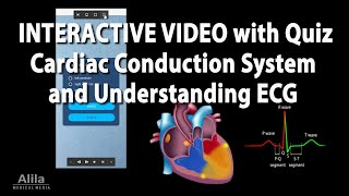
Cardiac Conduction System and Understanding ECG/EKG - Updated 2024, Animation

HTML-код
- Опубликовано: 31 май 2024
- Cardiovascular Physiology. Electrical Conduction Through the Heart and How Different Segments and Waves of the EKG (ECG) are Generated.
This is an updated version of our hugely popular video of the same name.
Purchase a license to download a non-watermarked version of this video here: www.alilamedicalmedia.com/-/g...
Purchase PDF (video text + images) here: www.alilamedicalmedia.com/-/g...
Check out our new Alila Academy - complete video courses with quizzes, PDFs, and downloadable images, here: www.alilaacademy.com/courses/
Join this channel to get access to member-only videos and other perks:
/ @alilamedicalmedia
©Alila Medical Media. All rights reserved.
Voice by Marty Henne
All images/videos by Alila Medical Media are for information purposes ONLY and are NOT intended to replace professional medical advice, diagnosis or treatment.
The cardiac conduction system consists of the following components:
- The sinoatrial node, or SA node, located in the right atrium near the entrance of the superior vena cava. This is the natural pacemaker of the heart. It initiates all heartbeat and determines heart rate. Electrical impulses from the SA node spread throughout both atria and stimulate them to contract.
- The atrioventricular node, or AV node, located on the other side of the right atrium, near the AV valve. The AV node serves as the electrical gateway to the ventricles. It delays the passage of electrical impulses to the ventricles, to ensure that the atria have ejected all the blood into the ventricles before the ventricles contract.
- The AV node receives signals from the SA node and passes them onto the atrioventricular bundle - AV bundle or bundle of His.
- This bundle is divided into right and left bundle branches which conduct the impulses toward the apex of the heart. The signals are then passed onto millions of Purkinje fibers and spread throughout the ventricles.
Electrical activities of the heart can be recorded in the form of an electrocardiogram, ECG or EKG. An ECG is a composite recording of all the action potentials produced by the nodes and the cells of the heart. Each wave or segment of the ECG corresponds to a certain event of the cardiac electrical cycle:
- When the SA node fires, electrical signals spread throughout the atria and cause them to depolarize. This is represented by the P wave on the ECG. Atrial contraction, or atrial systole, starts about 100 milliseconds after the P wave begins.
- The PR interval, which measures from the start of P wave to the start of QRS complex, represents the time between atrial depolarization and ventricular depolarization, and reflects the conduction through the AV node.
- The QRS complex represents ventricular depolarization:
- Q wave corresponds to depolarization of the interventricular septum.
- R wave is produced by depolarization of the main mass of the ventricles.
- S wave represents the last phase of ventricular depolarization toward the base of the heart.
- Atrial repolarization also occurs during this time but the signal is obscured by the large QRS complex.
- The ST segment reflects the plateau of action potentials in the ventricles. This is when the ventricles contract and pump blood.
- The T wave represents ventricular repolarization immediately before ventricular relaxation, or ventricular diastole. Because the rate of repolarization is slightly different for the 3 layers of the heart wall, the peak of T wave reflects repolarization of epicardial cells, while the end of T wave corresponds with repolarization of mid-myocardial cells.
The cycle repeats itself with every heartbeat.


Love our videos? Check out our new courses made entirely with videos like this (no watermark, no ads): www.alilaacademy.com/
NEW: Now you can also get access to our courses with our RUclips channel membership (Academy Access levels)
Got my step 1 result ... ur channel helped me a lot thank u so much ... btw i passed ❤
AMZING YALL ARE THE BEST SAVING FUTURE DOC❤❤❤❤❤
Penjelasannya jelas, terima kasih😊
Thanks .
I saw the title and immediately thought of the protagonist from Katawa Shoujo, whose problem *is* a heart conduction disorder.
Pls sir tell me about differnce between ST and QT segment
This may help you , ST segment is starting from J point ( jonction ) after QRS complex to the start of T wave . QT segment englobes both the ventriclar depolarization and repolarization it starts from the beginning of QRS complex to the last point of T wave .
🙏💯👍