Dr.Minarcik, I just wanted to say that you are saving students in gdansk medical university in Poland :) today is the final practical in about and hour and a half. These videos are a masterpiece. Thank you so much.
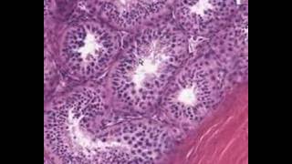
theca interna has a receptor for LH (luteinizing hormone) and makes progesterone and androgens (which are later made into estrogens) theca externa doesn't make diddly squat
wow...I found today your channel and I want to thank you because the videos are helping me a lot ! I am a medical student , and this year I started with histology and it seemed so strange , boring and hard to understand..the idea is that in one month I'm having the final exam and I'm so happy that I found this channel :D Thank you very much !Is really great what you are doing! greetings from Romania :D
You know that the oocito stages have never quite stuck with me up till today; and therfore MUCHISIMAS GRACIAS for the developmental outlook it realy put it into perspective!!!!
as i learn, secondary follicule has theca layers, zona pellucida and small antrums, but not one big antrum, . And the follicule that we call graaf has one big antrum + kumulus ooforus + corona radiata + theca layers +zona pellucida etc. Also primer follicule has zona pellucida and granulosa cells too. We seperate primer follicule as early and late phases. On early phase, we can see one layer cuboidal cells; on late phase we can see multilayer granulosa cells, no antrum.
Thank you so much, especially for offering one of the very few opportunities of a free of cost tutoring! Very, very appreciated. :) Awesome overview for me before diving deeper into the topic!
Maybe it is a different system, but in my Universitie's exams at the secondary oocyte you also have to explain about the Theca cells.. Great video however, thank you.
By the way.. I wanna thank you personnaly cuz It has help me trough my histology lab and I think this is the best way to study at home with the help of our books, I wanted also to say that the cells that are aroud the ovocyte (in the folliculary liquid can be called CORANA RADIATA and that the CUMULUS OOPHORUS are the cells that "attached" the ovocyte to the ZONA GRANULOSA.
not sure if "memorize" is going to help, remembering that ALL shades of blue, red, and purple will have tremendous variations in H&E with different labs, days, and techs i think memorizing colors is best for NON H&E stains like trichrome (collagen green), prussian blue (hemosiderin dark green) and PAS (bright red)....thise are the colors which do NOT vary
Don't know if you're alive or not But please keep making these videos What i learn in my med school is a lot of bullshit wasting time with egoistic teachers Once again thank you very much!!
Can you possibly email me a class photo if you have one, sometimes I have to be reminded the reason why I work so hard to do all this stuff! Kindly, Dr. Minarcik
You are the best professor on planet! So relaxed, and sharing the right amount of information I wish you could come to our university and show them how it's done!
Cumulus Oophorus as far as I know is at the base of the oocyte, away from the antrum, while the rest of the granulosa cells surrounding the oocyte go by the name of Corona Radiata. Please correct me if I'm wrong.
On the labeled slides from university of iowa they show your secondary follicles as primary follicles and your de graaf follicle as a secondary one.I'm pretty sure they're wrong.. aren't they?
i really like the way you explain the slides, very easy to understand ! i have just a question about the theca follicle. where is it ? shouldn´t it sorround the follicle with an internal and external layer ? or am i just confused
Hey! Congrats on the whole shotgun histology series, it's awesome. I just have one question. I've read that secondary follicles are the ones with the formed antrum, while the graafian follicles are the ones with a lose oocyte in the middle of the follicle... is this true? which is which? thanks!
Hi sir, I've been watching your videos for a while now, but couldn't figure it out.. Which program do you use? How do you get those images? Is there a free online photo database with these?
Hi Professor... In a biopsy of an ovary of a postmenopausal woman, how can I say it's an ovary when I don't see any follicles? I simply can't tell wether something is an ovary just by looking at the stroma, can I? Thank you!
Thank you! I've been looking at my universities biopsies of different ovarial tumors of postmenopausal ovaries and grew desperate trying to find any semblance of normal tissue. Thank you again, and thank you for all your videos!
When you shoot a shotgun, you can't miss a thing. When you see a slide you can't miss the name of anything. I believed this was explained in the first video.


Dr.Minarcik, I just wanted to say that you are saving students in gdansk medical university in Poland :) today is the final practical in about and hour and a half. These videos are a masterpiece. Thank you so much.
theca interna has a receptor for LH (luteinizing hormone) and makes progesterone and androgens (which are later made into estrogens)
theca externa doesn't make diddly squat
The second year med students from Cluj Napoca Romania thank you very much for putting so much effort to create these awesome videos.
Thank you SO much! Very helpful. I learned more in four minutes than I have in a week of class.
Light aiming at the flats ready to be deployed 😂
wow...I found today your channel and I want to thank you because the videos are helping me a lot ! I am a medical student , and this year I started with histology and it seemed so strange , boring and hard to understand..the idea is that in one month I'm having the final exam and I'm so happy that I found this channel :D Thank you very much !Is really great what you are doing! greetings from Romania :D
You know that the oocito stages have never quite stuck with me up till today; and therfore MUCHISIMAS GRACIAS for the developmental outlook it realy put it into perspective!!!!
as i learn, secondary follicule has theca layers, zona pellucida and small antrums, but not one big antrum, . And the follicule that we call graaf has one big antrum + kumulus ooforus + corona radiata + theca layers +zona pellucida etc. Also primer follicule has zona pellucida and granulosa cells too. We seperate primer follicule as early and late phases. On early phase, we can see one layer cuboidal cells; on late phase we can see multilayer granulosa cells, no antrum.
Thank You - I am glad you are generously sharing your knowledge here. Your hard work and talent are appreciated!
The virtual slides are from the Univ of Iowa website
Thank you sir....i have grown by learning thru your videos
Thank you so much, especially for offering one of the very few opportunities of a free of cost tutoring! Very, very appreciated. :)
Awesome overview for me before diving deeper into the topic!
Did you mean to say that the "oogonia" (rather than "oocytes") turn into primordial follicles, since technically every stage contains oocytes?
Sir, Thank you so much for the explaination! You saved my test.
Amazing explanation!
Thank you so much for sharing all of your knowledge! You make histology easier and interesting! I love all of your videos.
this is a piece of art.
Thank-you sir.
Still great!🤩
Excellent video, slides and explanation. Thanks
Maybe it is a different system, but in my Universitie's exams at the secondary oocyte you also have to explain about the Theca cells.. Great video however, thank you.
Thank you for the nice presentation and easy to understand slides.
Succes la examenul de histologie! Coleg de medicina an 2 UMF Bucuresti
finally some one who wants to teach!!! life saver
By the way.. I wanna thank you personnaly cuz It has help me trough my histology lab and I think this is the best way to study at home with the help of our books,
I wanted also to say that the cells that are aroud the ovocyte (in the folliculary liquid can be called CORANA RADIATA and that the CUMULUS OOPHORUS are the cells that "attached" the ovocyte to the ZONA GRANULOSA.
Great slides and great explanation 👍 thanks a ton!
Achei Muito didático este video sobre a histologia ovariana e desenvolvimento folicular. Parabéns ....
U r awesome shak...u made my day!
not sure if "memorize" is going to help, remembering that ALL shades of blue, red, and purple will have tremendous variations in H&E with different labs, days, and techs
i think memorizing colors is best for NON H&E stains like trichrome (collagen green), prussian blue (hemosiderin dark green) and PAS (bright red)....thise are the colors which do NOT vary
Don't know if you're alive or not
But please keep making these videos
What i learn in my med school is a lot of bullshit wasting time with egoistic teachers
Once again thank you very much!!
Last time I checked, i was still alive!
@@WashingtonDeceit Man thanks for these videos. Super helpful and I love em! Helps a ton in med school
Can you possibly email me a class photo if you have one, sometimes I have to be reminded the reason why I work so hard to do all this stuff!
Kindly,
Dr. Minarcik
You are the best professor on planet! So relaxed, and sharing the right amount of information
I wish you could come to our university and show them how it's done!
Excellent explation short and crisppp+!
You are great teacher.
Cumulus Oophorus as far as I know is at the base of the oocyte, away from the antrum, while the rest of the granulosa cells surrounding the oocyte go by the name of Corona Radiata. Please correct me if I'm wrong.
On the labeled slides from university of iowa they show your secondary follicles as primary follicles and your de graaf follicle as a secondary one.I'm pretty sure they're wrong.. aren't they?
Great explaination! Thank you
i really like the way you explain the slides, very easy to understand !
i have just a question about the theca follicle. where is it ? shouldn´t it sorround the follicle with an internal and external layer ? or am i just confused
most likely you are correct, i dont put labels on my movies, i just point and talk, sometimes i make boo-boos
Hey! Congrats on the whole shotgun histology series, it's awesome. I just have one question. I've read that secondary follicles are the ones with the formed antrum, while the graafian follicles are the ones with a lose oocyte in the middle of the follicle... is this true? which is which? thanks!
Wasnt the cartridges found on the grounds?
Hi sir,
I've been watching your videos for a while now, but couldn't figure it out.. Which program do you use? How do you get those images? Is there a free online photo database with these?
dude that why i do them
cuz i know you love them!
wdc
u made my day!
great videos!
Hi!!! i wanted to ask u sth about the theca!!!
when are the theca interna and externa produce???
Thank u professor
Hi Professor... In a biopsy of an ovary of a postmenopausal woman, how can I say it's an ovary when I don't see any follicles? I simply can't tell wether something is an ovary just by looking at the stroma, can I?
Thank you!
You are correct, might be impossible if it's a small biopsy, small biopsies are almost never done on ovaries however.
Thank you! I've been looking at my universities biopsies of different ovarial tumors of postmenopausal ovaries and grew desperate trying to find any semblance of normal tissue.
Thank you again, and thank you for all your videos!
Thanks a lot ....🤘
The primary follicles will also have the zona pollecida ,
Thank you so much!!
Now I understand feelings. Strokeitmuself.
the med students in Anguilla thank you so for your help
Thanks to you!
thank you so much!
Thank you :)
why is it called shotgun tho
When you shoot a shotgun, you can't miss a thing. When you see a slide you can't miss the name of anything. I believed this was explained in the first video.
Merci bro. You too.
AHHHHHHHHHHH YES SAN DIEGO STATE GO SAN DIEGO STATE
believe whatever your own school tells you because that it what those suckers might put on the test....plus...ahem....i might be wrong too...hehe
John Black please email me a class photo if you have one easily available, thank you very much for the effort! Dr. Minarcik minarcik@gmail.com