ImageJ 101 For Every PhD with Image Data!
US
Войти
How to automate image processing in ImageJ with a single click (How to create a macro in ImageJ)
8:40
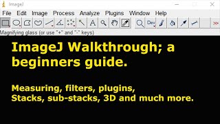
A beginners guide to ImageJ (and Fiji)
41:36
Getting The Most Out of Scientific Conferences
7:34
Gas Fruit Is The MOST OVERPOWERED Fruit.. (Blox Fruits)
10:37
Imagine Dragons - Take Me To The Beach (feat. Ado) (Official Lyric Video)
02:48
Demetrious Johnson Trains w/ KHABIB & ISLAM MAKHACHEV! | EXCLUSIVE FOOTAGE!
43:59
ImageJ 101 For Every PhD with Image Data!
PhDCoffeeTime
Подписаться
21 тыс.
Скачать
Готовим ссылку...
Просмотров 50 тыс.
1 100
0
Добавить в
Мой плейлист
Посмотреть позже
Поделиться
Поделиться
HTML-код
Размер видео:
1280 X 720
853 X 480
640 X 360
Показать панель управления
Автовоспроизведение
Автоповтор
Опубликовано: 7 фев 2025
Комментарии • 117
Следующие
Автовоспроизведение
8:40
How to automate image processing in ImageJ with a single click (How to create a macro in ImageJ)
Alicerita
Просмотров 17 тыс.
41:36
A beginners guide to ImageJ (and Fiji)
Craig Daly
Просмотров 94 тыс.
7:34
Getting The Most Out of Scientific Conferences
PhDCoffeeTime
Просмотров 5 тыс.
10:37
Gas Fruit Is The MOST OVERPOWERED Fruit.. (Blox Fruits)
FoltynPlays
Просмотров 871 тыс.
02:48
Imagine Dragons - Take Me To The Beach (feat. Ado) (Official Lyric Video)
ImagineDragonsVEVO
Просмотров 2 млн
43:59
Demetrious Johnson Trains w/ KHABIB & ISLAM MAKHACHEV! | EXCLUSIVE FOOTAGE!
Mighty
Просмотров 2,4 млн
03:21
Rio Da Yung OG - RIO FREE (Official Video)
Rio Da Yung OG
Просмотров 4,9 млн
10:44
Western Blotting | Beginner Data Interpretation Tutorial & Step-by-Step Protocol Explained
Emma Sandy
Просмотров 61 тыс.
23:36
ImageJ Analysis: Length Measurement, Area Measurement and Thresholding
SMS TechEdu
Просмотров 613 тыс.
9:34
Using ImageJ to measure cell number and cross-sectional area of confocal images
Dory Video
Просмотров 146 тыс.
11:01
How to Count Cells in 3D using ImageJ (Fiji)
MiketheMichael
Просмотров 45 тыс.
44:20
Intro to ImageJ/Fiji
Harvard Center for Biological Imaging
Просмотров 119 тыс.
12:55
A Strange Map Projection (Euler Spiral) - Numberphile
Numberphile
Просмотров 1,3 млн
18:32
How to read and take notes like a PhD - easy, fast, and efficient
Andy Stapleton
Просмотров 441 тыс.
24:42
Bioimage Analysis - Christian Tischer (EMBL)
iBiology Techniques
Просмотров 12 тыс.
00:49
Avoid The Person Challenge 😱
Zhong
Просмотров 7 млн
00:31
Лечение болезни Паркинсона
Новостной Гусь
Просмотров 946 тыс.
00:20
Feeling someone’s watching you👩🏻💻 #VictoriaPfeifer
Victoria Pfeifer
Просмотров 14 млн
1:04:49
Трамп, доллар и нефть: Экономист Игорь Липсиц о том, что ждет экономику России и Казахстана?
Арманжан Байтасов
Просмотров 96 тыс.
45:38
БЕЗНАКАЗАННЫЙ ТРЕШ-САЛОН ХАМИТ КЛИЕНТУ 😰/ Треш-обзор салона красоты в Москве
MOLOTOWA
Просмотров 190 тыс.
00:15
САМОЕ ПОЛЕЗНОЕ МАТЕМАТИЧЕСКОЕ УРАВНЕНИЕ #Shorts #Глент
ГЛЕНТ
Просмотров 2 млн
14:41
Куда делись яйца в США ? И чем теперь завтракать?
АМЕРИКА Наизнанку
Просмотров 136 тыс.
00:59
Необычный румтур 🤭 #сашаспилберг
Sasha Spilberg
Просмотров 577 тыс.