Picture tests in histology reproductive system - male

HTML-код
- Опубликовано: 12 дек 2024
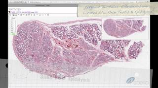
- After completion of this session it is expected that the students will be able to identify, locate and describe the histological features of:
Male genital organs:
Seminiferous tubules: Spermatogenic cells: various stages of spermatogenesis and spermiogenesis: spermatogonia, spermatocytes and spermatogonia; Non-spermatogenic cells: Sertoli cells; Myofibroblasts and fibroblasts in the supporting tissue; Outline the structural changes in spermiogenesis; Sertoli cells: Describe their shape and position; Discuss their role in the formation of the blood-testis barrier; Describe the shape of the spermatozoon and its parts: head, neck and tail; Locate Leydig cells and outline their function.
Epididymis
Outline its function and relate structure to function: role of muscular layer and stereocilia; Describe the layers in its wall: epithelium and muscular layer.
Ductus (vas) deferens
Outline its function and relate structure to function: role of muscular layer and stereocilia; Describe the layers in its wall: epithelium and muscular layer.
Seminal vesicles
Describe the layers in its wall: epithelium and muscular layer and relate that to function; Identify the honeycombed appearance of its lumen and the foamy cells of mucosa; Give reason why it may contain spermatozoa.
Some images were cited in histology guide a virtual histology laboratory www.histologygu...
Presented and edited by Akram Jaffar, PhD.
This video and its channel are supported by "Human Anatomy Education" Page on Facebook / anatomyeducation


Thank you so much for making these vids, it helps me a lot considering I've been struggling gravely in Histology.
Thank you! These exercises are helping me to study for my Histology tests!
King of Anatomy and Histology
best of the best mashallah
thanks alot
Thanks! glad it was helpful.
Videos ease the work of interpretation
Very much appreciated Dr. J
You are very welcome
Amazing video. Very helpful
thanks a lot for videos. amazing explanations.
Thank you.
Welcome!
Thanks alot doctor
Always welcome
Legend!!
You're welcome!
thanks for this ...
+pei broker thank you for the interest in the channel.