Please compile all your videos systematically as parts and present as normal studies as well as important and less common anomalies that we must identify in that mode..for example how to perform m mode ,what normally we should see,what important information we should look for and what are abarant findings
You ignored to correct the angle of insonation. It also looks like flow is well beyond the 60-degree limit of angle correction. So the velocities you measured are about 2x higher than the correct values.
@Danger Manilla Thank you for your comments. Although there has NOT been any scientific data to showing risk to the fetus with the use of ultrasound the principle of ALARA (as low as reasonably achievable) should be recognized when performing ultrasound on a fetus. To do so with ultrasound in pregnancy ultrasound one should limit the time to perform the ultrasound as much as possible and I'd recommend you use M-Mode instead of doppler to document fetal heart rate. ruclips.net/video/v4Vn27dMAEw/видео.html Here is an article publish by WHO regarding biological effects of ultrasound on a fetus. doi.org/10.1002/uog.6328
I watch one of the video and now i am confuse is we are getting wave form after identifying and freezing the image and then place the SV on the point we are looking for ?
While you are in standard scanning mode (B mode or 2 D Mode) you will NOT freeze the image. Push the button for Doppler (some machines with saw Pulse Wave, PW or similar) and place the gate over the area of interest. After this, depending on the machine, you will push the update button or again push the doppler button. This will bring a screen tracing up like you see in the video. This will be the example of you pulse wave doppler.
Thanks so much sir. Iam using esaote MyLab30 sir kindly guide me how i will get waveform doppler after locating the artery and placing the gate as you mention above that different machine has different option. Once again thank you Sir.


Great explanation! Always handy to watch a youtube explanation after reading the chapter in the book. Thank you.
Please compile all your videos systematically as parts and present as normal studies as well as important and less common anomalies that we must identify in that mode..for example how to perform m mode ,what normally we should see,what important information we should look for and what are abarant findings
thanks a lot
You ignored to correct the angle of insonation. It also looks like flow is well beyond the 60-degree limit of angle correction. So the velocities you measured are about 2x higher than the correct values.
Yep your are correct. The purpose of this video was to explain the concept of pulse wave doppler not to discuss angle of insonation.
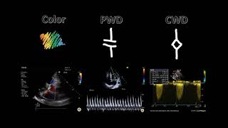
Hey ....where is you continuous wave Doppler video
Nice
You try to give the video more brightness it will be great if you do
Hi Sir. Thanks for the great explanation. Would you be doing a video about continuous wave Doppler as well?
Currently I don't have any plans to do a video on continuous doppler since it is not typically used in point of care ultrasound.
@Danger Manilla Thank you for your comments. Although there has NOT been any scientific data to showing risk to the fetus with the use of ultrasound the principle of ALARA (as low as reasonably achievable) should be recognized when performing ultrasound on a fetus.
To do so with ultrasound in pregnancy ultrasound one should limit the time to perform the ultrasound as much as possible and I'd recommend you use M-Mode instead of doppler to document fetal heart rate. ruclips.net/video/v4Vn27dMAEw/видео.html
Here is an article publish by WHO regarding biological effects of ultrasound on a fetus. doi.org/10.1002/uog.6328
@Danger Manilla Could you provide references for the studies you refer to?
Hello sir, do you have lecture on continuous wave doppler? Thank you!
I watch one of the video and now i am confuse is we are getting wave form after identifying and freezing the image and then place the SV on the point we are looking for ?
While you are in standard scanning mode (B mode or 2 D Mode) you will NOT freeze the image. Push the button for Doppler (some machines with saw Pulse Wave, PW or similar) and place the gate over the area of interest. After this, depending on the machine, you will push the update button or again push the doppler button. This will bring a screen tracing up like you see in the video. This will be the example of you pulse wave doppler.
Thanks so much sir. Iam using esaote MyLab30 sir kindly guide me how i will get waveform doppler after locating the artery and placing the gate as you mention above that different machine has different option. Once again thank you Sir.
Sir kindly give me your email address so i can send images for further discussion. Thank you
pocusgeek@gmail.com
How to measure heart rate by using this Doppler freeze image
Sir great video. What is the Doppler angle, it's significance, how and when to put it. Pls help.
Insonation angle for best velocity measurement align probe in line with flow 0 degree. upto 20 degrees acceptable
thanks alot