Fate Map during Gastrulation - 3D Human Embryology - Third Week Embryology

HTML-код
- Опубликовано: 28 июл 2024
- As we had discussed in the last lecture of somitogenesis and changes in intraembryonic mesoderm, that the intraembryonic mesoderm is not a single, homogenous undifferentiated layer. It is, instead, made up of different parts viz., Notochord or axial mesoderm, paraxial mesoderm, intermediate mesoderm, and lateral plate mesoderm.
This process called gastrulation, during which there is the formation of different types of mesoderm occurs, is not a random, haphazard process but rather a nicely coordinated process.
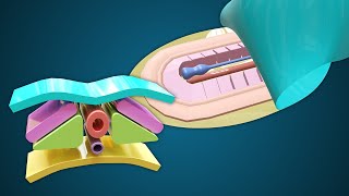
As we had seen in the lecture of gastrulation, the cells from the epiblast layer ingress downwards through the primitive streak and primitive node to first replace the hypoblast layer and then form different components of intraembryonic mesoderm between the two layers.
The primitive streak, along with the primitive node [Hensen’s node], is the real guiding star during gastrulation.
The fate of these ingressing cells is determined by the location or part of primitive streak and node from which they ingress downward. Let me explain how.
The cells that ingress downward from the primitive or Hansen’s node will form the notochord and prechordal plate.
Cells that ingress down from the cranial most part and caudo-lateral part of primitive node will form the paraxial mesoderm.
Cells that ingress from the midstreak will form the intermediate mesoderm
Cells that ingress from the caudal part of strak will form the lateral plate mesoderm.
Finally, the cells that ingress from the caudal most end of the streak will contribute slightly to the extraembryonic mesoderm.
Remember that most of the extraembryonic mesoderm is thought to have been originated, during the second week, from the hypoblast cells. However, some contribution also done by these ingressing cells during the gastrulation in third week of development.
What is extraembryonic mesoderm by the way. As you must be knowing the ectoderm continues with amnion and endoderm layer continues with the yolk sac. So, baby is provided with these two fluids filled balloons. Outside these two balloons and the embryo proper, whole this structure is plastered outside with a layer of extraembryonic mesoderm.
Website: www.medicovisual.com
Email: draizaz@medicovisual.com


![Gastrulation - Human Embryology - 3rd Week [Animated] - MedicoVisual](http://i.ytimg.com/vi/0n_Ev8DbQgY/mqdefault.jpg)


None of the video is more good than yours when it comes to embryology!
respect for India people and doctors 🇮🇳 ❤💚👍👍👍👍👍
Why are you this awesome?!!! I wish I could like and subscribe to your channel more than once. Thank you so much for your videos.
Indeed you and your animated vedios are live saving ❤
Jazak Allah sir
Thank you sooo much
Very good 3D graphic presentation of developmental Embriology.
Thanks
Thank you for this well explained video
You are welcome!
This 3d structure is so eatable. Thank you so much for the video.
Thank you so very much for watching (Finally!)
@@MedicoVisual 😌😌
thank u for the video! greetings from argentina :)
You are very welcome.
Nice animation. loved it . the growth of embryo is cephalocaudal but here the cells are going in opposite direction. . is there any specific reason?
Thank you. As far I know, the cell ingress downward and spread cranially (as in animation). Please correct me if I am wrong.
Amazing video sir 🤠🤠🤠.
Sir ,Upload more animated videos on embryology .
Thank you so much. I am working on more of such visual lectures. Stay Blessed
Does these mesoderm layers exist above the buccopharangeal membrane? What forms the septum transversum?
Buccopharyngeal membrane (or Oropharyngeal membrane) is an oval shaped double-layered membrane present at the cranial end of the embryo that contains ectoderm and endoderm with little to no mesoderm between them. (Watch this video: ruclips.net/video/jTymhPysNOM/видео.html)
Spetum Transversum has been discussed in detail in Heart Tube lecture. Watch here: ruclips.net/video/tcM7DOSoabI/видео.html
Please upload video development of placenta
Visual lectures on "Development of Placenta" have not yet been recorded. They will be recorded and uploaded within a week or so. Meanwhile watch this video for the basic overview of early development of placenta: ruclips.net/video/9YHPCvcoNFs/видео.html
Sir haven't you taught 4th week of development?
Not yet
@@MedicoVisual sir plllzzzzz make more videos of embryology lectures. Only you can teach embryology in the best possible manner🙏🙌
@@areebanadeem4988 Thank you so much. Trying my best
Thanks but please have sub i don't get the accent
But animation was great