*You are a saviour of embryology* *My Professors are too good in embryology but they neglected our embryology and histology lectures.* *But you are here to cope up with the deficiency* *Thanks a lot*
I think your way of teaching is unique and I'm glad I found this channel before falling into confusion.I like how you make little jokes to make it all so interesting
❤❤❤ Wow! It's amazing! More power to you for bringing such quality content! Now I can read this topic in Langman without any headache 😅! Thank you very much Sir!
Urogenital System Embryology is already available for a small fee at www.medicovisual.com/courses/premium-urogenital-system-3d-masterclass Do not hesitate to contact me if you have any questions or concerns. WhatsApp: +923037241030 Email: draizaz@medicovisual.com
VEGF is secreted by the surrounding mesodermal cells.. Suppose if blood islands are formed within the extraembryonic splachnic mesoderm, the process of blood vessels formation in these blood islands is stimulated by the VEGF secreted by the surrounding mesodermal cells of extraembryonic splanchnic mesoderm.
@@AlKubaisi96 No problem. Here you go: Langman's Medical Embryology. Pg. 86 (Under the heading of "Molecular Regulation of Blood vessel formation") books.google.com.pk/books?id=UTjvDwAAQBAJ&pg=PA86&dq=Vegf+is+secreted+by+surrouding+mesoderm+embryology&hl=en&sa=X&ved=2ahUKEwjT95b-1MP6AhV2h_0HHVvqB7QQ6AF6BAgCEAM#v=onepage&q=Vegf%20is%20secreted%20by%20surrouding%20mesoderm%20embryology&f=false
Yes. Vitelline Blood vessels are formed from the extraembryonic mesoderm overlying the yolk sac. These vitelline vessels will form the celiac trunk, superior mesenteric artery and vein and inferior mesenteric artery and vein in the adults. I have discussed this in detail in the Cardiovascular Embryology course (paid) on MedicoVisual.com
I am trying to upload as often as possible. However , due to intensity of effort in these videos, its impossible to do so. I don't have a team. I am the sole person to research the topic first, then make the graphics of it and record it myself.
Sir, the extra embryonic mesoderm is divided into 2 layers. 1st is splanco mesoderm then is mesodermal cellom and then is somatic layer of mesoderm ... so most upper one is extra embryonic somatopleuric mesoderm , not the splanic one. So now from where blood islands Start forming ? If blood islands come from upper most then it can not be spalnic cause above it is cellom then somatic layer. So plz clear Kindly clear it ? Answer must. Thank you
Mesoderm overlying the yolk sac is extraembryonic splanchnic mesoderm while the one that overlies amnion is somatic layer. Blood island first form in the extraembryonic splanchnic layer overlying the yolk sac.
@@medicalscience1528 No dear. Somatopleuric mesoderm does not cover the yolk sac. I know some USMLE resources have wrongly written it otherwise. (This confused me as well when I was creating the lecture series of 2nd week of development). However almost every major textbook of embryology including Langman, Larsens, Bruce Carlson, Keith Moore and Inderbir Singh clearly mentions that somatopleuric mesoderm covers the amnion and splanchnopleuric mesoderm covers the yolk sac cavity. This view is quite logical as well. If you study the lateral folding and craniocaudal folding, you will realize that the body wall is formed by the intraembryonic somatopleuric mesoderm (that is continous with extraembryonic somatopleuric mesoderm covering the amnion). The word "Somato" means "pertaining to the body wall". So it is very logical to call the mesoderm covering amnion as somatopleuric mesoderm. Opposite to that, if you look at the yolk sac and the endoderm, they will form the viscera of GIT and thus are plastered with splanchnopleuric (also called visceral) mesoderm. Just think about it, how can "somatic" mesoderm cover the viscera?
1. Developing Human 10th edition by Keith Moore 2. Larsens Human Embryology by Gary C. Schoenwolf, Steven B. Bleyl, Philip R. Brauer, Philippa H. Francis-West 3. Langman’s Medical Embryology 4. Inderbir Singh's Human Embryology 11th Edition 5. Embryology an illustrated colour text by B S Mitchell R Sharma Robert Britton 6. Human Embryology Developmental Biology by Bruce M. Carlson 6th ed 7. Human Embryology Made Easy by Abdul Hamid Rana 8. Clinical Embryology Robert Carachi 9. Embryology by Robert E Coalson 10. Human Embryology Prenatal Development of Form and Function by W. J. Hamilton 11. Developmental biology by Michael J. F. Barresi, Scott F. Gilbert
You can always contact me if you cannot afford any of my course. I will definitely accommodate you. 😊 I want every student to benefit from my lectures. However, it is difficult to sustain this project without paid courses. I hope you can understand. You can always text me on WhatsApp at +923037241030 or Email at draizaz@medicovisual.com
I was quite enjoying the content until you starting bringing in quite unprofessional examples. There wasn’t any need of them. Please remain professional when delivering education. Many people look up to you; you shouldn’t propagate the use of such low jokes. Thanks.
@@nirgunkaur3161 These were to impart some interest and keep the audience hooked and also to help the students remember. I am sorry that you didn't like it. I will try to refrain from using such examples as much as possible in the upcoming lectures.
@@karanteraiya1768 Also, my intention by making such jokes is to improve long-term retention. I still remember I watched one of the Dr. Najeeb's lectures, years ago. He said, sixth cranial nerve is sex nerve. It goes inside the room (cavity of cavernous sinus) and has intimate relationship with carotid artery.


What a passionate teacher you are. Thank you so much!
Give this man a nobel prize , because he's saving millions of lives from confusion
Yes
*You are a saviour of embryology*
*My Professors are too good in embryology but they neglected our embryology and histology lectures.*
*But you are here to cope up with the deficiency*
*Thanks a lot*
Sir, you are a life saver, my lecture notes now make sense. Thank you
I think your way of teaching is unique and I'm glad I found this channel before falling into confusion.I like how you make little jokes to make it all so interesting
You are special💯 ,we need more from you pls do more and save from this bulky medical life .I don't know how to express my thanks , God bless you.🤗👌👌👌💯
This really helps me understand a crucial part of my research project, thank you so much!
❤❤❤ Wow! It's amazing! More power to you for bringing such quality content! Now I can read this topic in Langman without any headache 😅! Thank you very much Sir!
Thank you so much for giving us exactly what we missing, can't thank you enough sir🙏🙂
Jazak Allah khairan kaseera
I wish, I were a real life student of you..
Thank you sir..
Very beautifully explained prrsentaion at this stage of development of embryo
Thanks
omg such informative video deep to the details you are the best thank you very much
Waiting eagerly
thanks sir for these wonderful lectures sir pls upload whole mbbs first year embryology on youtube
can you please make a video about cardiogenesis? Thank you for all the amazing work!
Will make it soon
Thank you so much! So necessary, both funny and interesting
Sir ur videos r really helpful..plz upload a video on the development of genital system
Urogenital System Embryology is already available for a small fee at www.medicovisual.com/courses/premium-urogenital-system-3d-masterclass
Do not hesitate to contact me if you have any questions or concerns.
WhatsApp: +923037241030
Email: draizaz@medicovisual.com
Great explanation good job
Very well explained
Thank you so much for your videos! SO helpful!! What app do yo use to visualize those 3D structures??
You are most welcome. I use Blender 3D
Are the endothelial precursor cells the same as angioblasts?
Awesome explanation 👍
Thank you 🙂
both processes are controlled by VEGF though receptors are different fig 6.14 pg 86 langmann
Thank you for clarifying.
Excellent Work
Many thanks
Amazing lecture sooooo goood thanks♥️
you are as funny as accurate, thank you sir, a pleasure to attend your lecture!
Sir, I have 1 more question ... can you please explain from where VEGF are released ? Who release these factors ?
VEGF is secreted by the surrounding mesodermal cells.. Suppose if blood islands are formed within the extraembryonic splachnic mesoderm, the process of blood vessels formation in these blood islands is stimulated by the VEGF secreted by the surrounding mesodermal cells of extraembryonic splanchnic mesoderm.
@@MedicoVisual Thank you so much sir , you know these tiny missing concepts always trigger if not cleared ... I really appreciate your reply back.
@@medicalscience1528 You are always welcome.
Hi thanks for the great videos, can I ask for the sources and origin of this information. I need to read more
@@AlKubaisi96 No problem. Here you go: Langman's Medical Embryology. Pg. 86 (Under the heading of "Molecular Regulation of Blood vessel formation") books.google.com.pk/books?id=UTjvDwAAQBAJ&pg=PA86&dq=Vegf+is+secreted+by+surrouding+mesoderm+embryology&hl=en&sa=X&ved=2ahUKEwjT95b-1MP6AhV2h_0HHVvqB7QQ6AF6BAgCEAM#v=onepage&q=Vegf%20is%20secreted%20by%20surrouding%20mesoderm%20embryology&f=false
Sir sorry to say again but sir could you upload cardiac embryology becoz I have lots of confusion please sir am eagerly waiting
No need to be sorry. You are most welcome to comment here. Out of the whole CVS Embryology, what topic(s) you find most confusing ?
finding it difficult to visualise the heart tubes fusing sir
@@77ranjanis62 Noted. I will do it as soon as possible.
Thank you so much!!!!!
Excellent 👌👌
Sir , is thi vessel at eem take part in any big vessel formation?
Yes. Vitelline Blood vessels are formed from the extraembryonic mesoderm overlying the yolk sac. These vitelline vessels will form the celiac trunk, superior mesenteric artery and vein and inferior mesenteric artery and vein in the adults. I have discussed this in detail in the Cardiovascular Embryology course (paid) on MedicoVisual.com
Thank you so much sir for your kind help🙏🙂
Extraordinary 👌👌👌👍👍👍
Thanks a lot
Amazing 👏
Thanks a lot sir ❤
Nice explanation 👌🔥
Thanks 🙂
Now I know.
Asante sana
please upload more often
I am trying to upload as often as possible. However , due to intensity of effort in these videos, its impossible to do so. I don't have a team. I am the sole person to research the topic first, then make the graphics of it and record it myself.
@@MedicoVisual respects sir. after surfing through the whole of youtube , i got clarity only in your videos . keep going sir , truly helpful !
@@77ranjanis62 I am honoured 😊
Sir, the extra embryonic mesoderm is divided into 2 layers. 1st is splanco mesoderm then is mesodermal cellom and then is somatic layer of mesoderm ... so most upper one is extra embryonic somatopleuric mesoderm , not the splanic one. So now from where blood islands Start forming ? If blood islands come from upper most then it can not be spalnic cause above it is cellom then somatic layer. So plz clear
Kindly clear it ? Answer must. Thank you
Mesoderm overlying the yolk sac is extraembryonic splanchnic mesoderm while the one that overlies amnion is somatic layer. Blood island first form in the extraembryonic splanchnic layer overlying the yolk sac.
@@MedicoVisual Somato pleuric covers both amniotic and yolk sac . Isn't it ? It overlaps all around embryoblast except umbilical cord ... ??
@@medicalscience1528 No dear. Somatopleuric mesoderm does not cover the yolk sac. I know some USMLE resources have wrongly written it otherwise. (This confused me as well when I was creating the lecture series of 2nd week of development). However almost every major textbook of embryology including Langman, Larsens, Bruce Carlson, Keith Moore and Inderbir Singh clearly mentions that somatopleuric mesoderm covers the amnion and splanchnopleuric mesoderm covers the yolk sac cavity. This view is quite logical as well. If you study the lateral folding and craniocaudal folding, you will realize that the body wall is formed by the intraembryonic somatopleuric mesoderm (that is continous with extraembryonic somatopleuric mesoderm covering the amnion). The word "Somato" means "pertaining to the body wall". So it is very logical to call the mesoderm covering amnion as somatopleuric mesoderm. Opposite to that, if you look at the yolk sac and the endoderm, they will form the viscera of GIT and thus are plastered with splanchnopleuric (also called visceral) mesoderm. Just think about it, how can "somatic" mesoderm cover the viscera?
Great questions, by the way.
@@MedicoVisual Ok.
And thank you 🌻
well explained, thank you!🤓
really helpfull 👍
thank you 🙂
what is your bibliography?
1. Developing Human 10th edition by Keith Moore
2. Larsens Human Embryology by Gary C. Schoenwolf, Steven B. Bleyl, Philip R. Brauer, Philippa H. Francis-West
3. Langman’s Medical Embryology
4. Inderbir Singh's Human Embryology 11th Edition
5. Embryology an illustrated colour text by B S Mitchell R Sharma Robert Britton
6. Human Embryology Developmental Biology by Bruce M. Carlson 6th ed
7. Human Embryology Made Easy by Abdul Hamid Rana
8. Clinical Embryology Robert Carachi
9. Embryology by Robert E Coalson
10. Human Embryology Prenatal Development of Form and Function by W. J. Hamilton
11. Developmental biology by Michael J. F. Barresi, Scott F. Gilbert
Thankyou sir 🥺
Please just keep going, don't make your vedios not for free,there are a lot of students who can't afford😢,thanks ❤❤❤
You can always contact me if you cannot afford any of my course. I will definitely accommodate you. 😊 I want every student to benefit from my lectures. However, it is difficult to sustain this project without paid courses. I hope you can understand.
You can always text me on WhatsApp at +923037241030 or Email at draizaz@medicovisual.com
Just wow.....
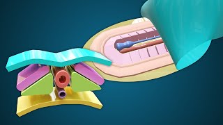
so that was the tip of endothelial cells following vgef i tought it was something else🤣🤣🤣🤣🤣
We are on the same page bro 🤣
10:20 LMAO
🤣
🫡🫡🫡
I was quite enjoying the content until you starting bringing in quite unprofessional examples. There wasn’t any need of them. Please remain professional when delivering education. Many people look up to you; you shouldn’t propagate the use of such low jokes. Thanks.
Which one specifically? Timestamp?
@@MedicoVisual 10:19
@@nirgunkaur3161 These were to impart some interest and keep the audience hooked and also to help the students remember. I am sorry that you didn't like it. I will try to refrain from using such examples as much as possible in the upcoming lectures.
I think that it's ok to make jokes as well it's just that way I think to keep audience in touch @@MedicoVisual
@@karanteraiya1768 Also, my intention by making such jokes is to improve long-term retention. I still remember I watched one of the Dr. Najeeb's lectures, years ago. He said, sixth cranial nerve is sex nerve. It goes inside the room (cavity of cavernous sinus) and has intimate relationship with carotid artery.
Of course that is a blood vessel don’t think of anything else 😂