This was SUPER interesting! Thank you for methodically going through each step in the process, while providing a very clear and detailed account. Your whole channel is pure gold 🏆
EXCELLENT explanation, and in perfectly understandable English. I've been a radiologist for 35 years, and you clearly know your stuff. I'm moving into the AI-imaging world; I look forward to more of your videos.
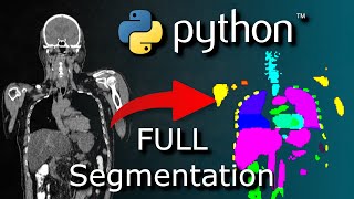
Awesome video! Thanks for showing the plotly thing at the end. I've been banging my head against the wall for a week trying to visualize MRI data in plotly without success.
Hey man, really appreciate what you’re doing. As a grad student myself, it’s really admirable that you teach us this stuff. Best wishes at school and life.
I've never worked with high level stuff like this, but I really enjoy your walkthroughs, because it helps me understand the thought process of performing complicated tasks such as this. I hope to do something this cool someday.
Hi, my jaw dropped on thie masterpiece, but I have one question: In the import file, you used a CT_scan.npy, wich means, and correct me if I'm wrong, that you transformed 200 layers of 1 exam that was .dcm in one single archive that is a group of arays .npy. Do you have any tutorial on how to do that or some link to how can I learn the way you did it? It would help a lot! Thanks!
Amazing video, thanks a lot. I'm trying to follow the steps, but when trying to download the file CT_scan.npy GitHub says the repository is over its data quota. Is there any other way of getting that file?
I couldn't find the "CT_scan.npy" file in the Data directory in your Github repo. Btw, do you have any tutorial on how to process the medical imaging files (dcm, MRI,...)
I like it so much when people take care about reproducibility of their research especially when it comes to computational science. Code should be supplementary material along with the data, and there shouldn't be "unnecessary hurdles on a way to reproducibility" (a citation from a Nature article as I recall). We have a technology for this like online repositories and code should also get doi numbers. Thanks for this video!
Thanks for the great video! I just have one question. I am trying to apply the same concept you discussed on CT scan of metallic parts (1024 x 1024). The challenge I have is that the code you used for finding boundaries and areas is not working properly due to having thin layers side by side. Do you have any suggestions as to how to fix this issue?
Excellent video. In my case, I have CT DICOM files that I’ve segmented using 3D slicer. I read those files in with sitk and need to work on processing them with labels the way you did here so that I can multiple the image x the labels and remove background nose before training into my model. Would love more videos along this line!
When cloning the project, I'm facing this issue ` Error downloading object: Data/CT_scan.npy (ee8b4e3): Smudge error: Error downloading Data/CT_scan.npy (ee8b4e370c617c0526360a761dd6051c16324b25496a705e39d65930a2511e6b): batch response: This repository is over its data quota. Account responsible for LFS bandwidth should purchase more data packs to restore access. `
Great video! I wonder how do you include more complex organs with lobes. For example, if you want to keep the trachea. I see that the lungs are kinda cut short on the bottom end because of the 3 biggest areas threshold. Would be cool to follow the diggestive system by using the center of mass function (at each layer, the center can shift a small amount).
Very interesting and useful video! I have a question: Is it possible to generate a 3d Image out of 2d Image Slices? I am currently trying it with plt.scatter, but theres always a distance between the single layers. Is it possible to concatenate those 2d slices and plot them as one element?
Interesting video, Thanks. There are many heuristics and parameters used in the video which may not work in the general case. So, I was wondering if you choose to look at slices that use the first 2 dimensions instead of the last 2 dimensions (different slices should be at different depths of the chest-back dimension), it might be easier to find out the location of the lungs and get rid of the table (the table should be less or more always at the same depth)? In any case, it must be assumed that this type of problem is more suitable for three-dimensional segmentation than the union of two-dimensional segmentations. Regardless, I would be very happy if you made a tutorial to image-processing libraries (2D) in the same style as you did for NumPy, SciPy & SymPy
thanks for such a nice walkthrough, I think it would have been much easier to make the code more modular, since you already wrote functions, you could have created a class to generate the image objects for you, but that is just a suggestion :) All the best.
@@MrPSolver Well... I'm still working with MATLAB. It has a nice IDE, no issues will mutual library dependencies and technically the code works faster than similar code in python, especially for the large datasets
Really clear and enriching tutorial ! Thank you very much ! I have a problem though at the last step, nothing displays with my HTML file, only the axis are showing up. Would you know if it's a problem of dimension in the data or type issue (my array in 'x', 'y', 'z' and 'value' are the same dimension and data type are int32).
The np.load method offered in the video tutorial does not open the .npy scan file. I was expecting an array in the file but can only see a sha and filesize... has anyone coded along with this and have a solution to my problem? Thanks
there is another video from another person for reading .dcm medical files . He shows you how to extract a .dcm file then convert it to numpy. Then you are ready to look at this again
@@ercankara3117 bulamadım onun yerine kendim bir kod yazdım. Dicom resimlerini dcmread le okuyup bir listeye attım. Daha sonra bu listeyi dicomun içinde bulunan ImagePositionPatient meta verisinin z-ekseni değerlerine göre sıraladım. Sıraladıktan sonra her bir pikseli HU değerlerine dönüştürüp 3D dizi oluşturdum.
Damn I thought it was "python segmentation fault tutorial( 2022)" I clicked wondering how the heck you billy achieved that but now my Disappointment Is Immeasurable and my day is ruined.


This was SUPER interesting! Thank you for methodically going through each step in the process, while providing a very clear and detailed account. Your whole channel is pure gold 🏆
I totally agree!!!
@@vladi1475S filepath = r'../Data/CT_scan.npy' img = np.load(filepath) Bu dosyayı nereden indirebileceğim konusunda bana yardımcı olabilir misiniz?
filepath = r'../Data/CT_scan.npy' img = np.load(filepath) Bu dosyayı nereden indirebileceğim konusunda bana yardımcı olabilir misiniz?
@ercankara3117
13 dakika önce
filepath = r'../Data/CT_scan.npy'
img = np.load(filepath)
Can you help me where I can download this file?
EXCELLENT explanation, and in perfectly understandable English. I've been a radiologist for 35 years, and you clearly know your stuff. I'm moving into the AI-imaging world; I look forward to more of your videos.
Awesome video! Thanks for showing the plotly thing at the end. I've been banging my head against the wall for a week trying to visualize MRI data in plotly without success.
It's not perfect, but plotly can come in handy;
filepath = r'../Data/CT_scan.npy' img = np.load(filepath) Bu dosyayı nereden indirebileceğim konusunda bana yardımcı olabilir misiniz?
@@MrPSolver filepath = r'../Data/CT_scan.npy' img = np.load(filepath) Bu dosyayı nereden indirebileceğim konusunda bana yardımcı olabilir misiniz?
Hey man, really appreciate what you’re doing. As a grad student myself, it’s really admirable that you teach us this stuff. Best wishes at school and life.
I've never worked with high level stuff like this, but I really enjoy your walkthroughs, because it helps me understand the thought process of performing complicated tasks such as this. I hope to do something this cool someday.
filepath = r'../Data/CT_scan.npy' img = np.load(filepath) Bu dosyayı nereden indirebileceğim konusunda bana yardımcı olabilir misiniz?
Your medical physics videos are the best stuff ever
Seconded
@ercankara3117
13 dakika önce
filepath = r'../Data/CT_scan.npy'
img = np.load(filepath)
Can you help me where I can download this file?
Where can I get the '../Data/CT_scan.npy' file that you're using in cell 2? @Mr. P Solver
did u find it
@@abdulmahshooqka4374 did u find it
@@abdulmahshooqka4374 no bro
Thank you so much for these videos. I have started getting more involved in CT scans, and these videos have been very inspiring.
Hi, my jaw dropped on thie masterpiece, but I have one question:
In the import file, you used a CT_scan.npy, wich means, and correct me if I'm wrong, that you transformed 200 layers of 1 exam that was .dcm in one single archive that is a group of arays .npy.
Do you have any tutorial on how to do that or some link to how can I learn the way you did it? It would help a lot! Thanks!
Amazing video, thanks a lot. I'm trying to follow the steps, but when trying to download the file CT_scan.npy GitHub says the repository is over its data quota. Is there any other way of getting that file?
I couldn't find the "CT_scan.npy" file in the Data directory in your Github repo. Btw, do you have any tutorial on how to process the medical imaging files (dcm, MRI,...)
did you find it??
@@mervebulbul5099 did you find it??
@@mervebulbul5099 buldunuz mu acaba
This video is amazing, just as libraries for segmentation in python, skimage is OP.
I like it so much when people take care about reproducibility of their research especially when it comes to computational science. Code should be supplementary material along with the data, and there shouldn't be "unnecessary hurdles on a way to reproducibility" (a citation from a Nature article as I recall). We have a technology for this like online repositories and code should also get doi numbers.
Thanks for this video!
Learning python and libraries from your channel is fun 😇
This is exactly what I have wanted,
I cannot thank you more man!
Nice description 👍🏻
If i will take several images of lungs and want to integrate them and make one image than how I can integrate that several images?
Hi Mr P Solver, great tutorial! Where can we fint the '../Data/CT_scan.npy' file mentioned in the code?
Did you find it?
Thanks for the great video! I just have one question. I am trying to apply the same concept you discussed on CT scan of metallic parts (1024 x 1024). The challenge I have is that the code you used for finding boundaries and areas is not working properly due to having thin layers side by side. Do you have any suggestions as to how to fix this issue?
Very clear tutorial! Thanks a lot!!
Hi thank you its great...where can i find CT_Scan.npy
Excellent video. In my case, I have CT DICOM files that I’ve segmented using 3D slicer. I read those files in with sitk and need to work on processing them with labels the way you did here so that I can multiple the image x the labels and remove background nose before training into my model. Would love more videos along this line!
@ercankara3117
13 dakika önce
filepath = r'../Data/CT_scan.npy'
img = np.load(filepath)
Can you help me where I can download this file?
FANTASTIC!!!! Love the video!!!! Pls do more of these videos!!!! Thank you!!! :)
@ercankara3117
13 dakika önce
filepath = r'../Data/CT_scan.npy'
img = np.load(filepath)
Can you help me where I can download this file?
When cloning the project, I'm facing this issue
`
Error downloading object: Data/CT_scan.npy (ee8b4e3): Smudge error: Error downloading Data/CT_scan.npy (ee8b4e370c617c0526360a761dd6051c16324b25496a705e39d65930a2511e6b): batch response: This repository is over its data quota. Account responsible for LFS bandwidth should purchase more data packs to restore access.
`
Any clever tips how I can apply this but extract the heart only?
I can't find CT_scan.npy?
Thanks for the video! How would i go to do a similiar thing, but with a .las file?
Great video! I wonder how do you include more complex organs with lobes. For example, if you want to keep the trachea. I see that the lungs are kinda cut short on the bottom end because of the 3 biggest areas threshold. Would be cool to follow the diggestive system by using the center of mass function (at each layer, the center can shift a small amount).
Hi, great tutorial!...May I ask you why the html file doesn't open? Is there another way to show it?
problem when loading CT_scan.npy :
ValueError: Cannot load file containing pickled data when allow_pickle=False
me too
Very interesting and useful video! I have a question: Is it possible to generate a 3d Image out of 2d Image Slices? I am currently trying it with plt.scatter, but theres always a distance between the single layers. Is it possible to concatenate those 2d slices and plot them as one element?
Are the "CT_scans" archives a set of DICOM images?
did u find it
Thank you very much.
Where can i get ct_scan.pny
did u find it
Interesting video, Thanks.
There are many heuristics and parameters used in the video which may not work in the general case. So, I was wondering if you choose to look at slices that use the first 2 dimensions instead of the last 2 dimensions (different slices should be at different depths of the chest-back dimension), it might be easier to find out the location of the lungs and get rid of the table (the table should be less or more always at the same depth)?
In any case, it must be assumed that this type of problem is more suitable for three-dimensional segmentation than the union of two-dimensional segmentations.
Regardless, I would be very happy if you made a tutorial to image-processing libraries (2D) in the same style as you did for NumPy, SciPy & SymPy
@ercankara3117
13 dakika önce
filepath = r'../Data/CT_scan.npy'
img = np.load(filepath)
Can you help me where I can download this file?
can you help segmenting kidney from CT scan?
can you do a video on object detection on a biology image?
which python version use in this video?? Thank you
wow you're my savior, i need it ... thanks!
where can I get the dataset
I am not finding it
this was extremely helpful! and nice visuals too, what more could I ask for
Money...😅,
thanks for such a nice walkthrough, I think it would have been much easier to make the code more modular, since you already wrote functions, you could have created a class to generate the image objects for you, but that is just a suggestion :) All the best.
Fantastic!
And definitely cheaper than Image Processing Toolbox in MATLAB ;)
Who needs MATLAB anymore ;)
@@MrPSolver Well... I'm still working with MATLAB. It has a nice IDE, no issues will mutual library dependencies and technically the code works faster than similar code in python, especially for the large datasets
Fuck me silly this is a good quality video. The concrete example really helped me wrap my head around all the material.
filepath = r'../Data/CT_scan.npy'
img = np.load(filepath)
Can you help me where I can download this file?
Really clear and enriching tutorial ! Thank you very much !
I have a problem though at the last step, nothing displays with my HTML file, only the axis are showing up. Would you know if it's a problem of dimension in the data or type issue (my array in 'x', 'y', 'z' and 'value' are the same dimension and data type are int32).
@ercankara3117
13 dakika önce
filepath = r'../Data/CT_scan.npy'
img = np.load(filepath)
Can you help me where I can download this file?
I learned in another video how to access and display and ct photo of .dcm type. Now problem is how to convert it to numpy?
use np.array and then do sanity check on data by printing types()
This is not working, the Data Provided in the Git repository is showing OS Error.
well explained!
Great video.
Wow, super interesting. Thanks for sharing.
Filling in the holes, Data destruction
Hi do you think it is possible to do in 2D images?
yes it worked for me with small animals
God i love your videos
The np.load method offered in the video tutorial does not open the .npy scan file. I was expecting an array in the file but can only see a sha and filesize... has anyone coded along with this and have a solution to my problem? Thanks
The scan file is encrypted unless someone can advise otherwise... @Mr.PSolver could you advise where we might find a similar ct scan file?
Hello, Can anyone send me datafile or link to download it. Thanks 🙏
Why is the data deleted now?
can you please provide the link for filepath: ct_scan.npy file please?
there is another video from another person for reading .dcm medical files . He shows you how to extract a .dcm file then convert it to numpy. Then you are ready to look at this again
So could you show the link of this video?
@@inhibited44
Hi @@inhibited44 Which video that you tell about? Can you share the name or a link?
@@mervebulbul5099 hangi video acaba bulabiildiniz mi
@@ercankara3117 bulamadım onun yerine kendim bir kod yazdım. Dicom resimlerini dcmread le okuyup bir listeye attım. Daha sonra bu listeyi dicomun içinde bulunan ImagePositionPatient meta verisinin z-ekseni değerlerine göre sıraladım. Sıraladıktan sonra her bir pikseli HU değerlerine dönüştürüp 3D dizi oluşturdum.
Wouldn’t it had been Better to change view by having a (512, 263, 512)
superb
Saw your video! Amazing Tutorial, subscribed immediately!
This Code is actually not working, what so amazed did you saw in it?
Super 👍
It must be nice to be a Genius❤😅🎉
You are very clever, I want to be you student,
Damn I thought it was "python segmentation fault tutorial( 2022)" I clicked wondering how the heck you billy achieved that but now my Disappointment Is Immeasurable and my day is ruined.
Absolutely amazing! Outstanding presentation. I'd be grateful to have you involved in my project. @saucerdesigner