Ultrasound guided dynamic needle tip positioning in peripheral vein and artery cannulation

HTML-код
- Опубликовано: 8 июл 2024
- If you’d like to see the video of an actual patient, visit • Ultrasound guided peri... and view my video: “Ultrasound guided peripheral venous cannulation in an actual patient” .
******** Keyboard Shortcuts(PC) *************
If you feel that the movie is moving too fast, you can control the movie with your computer keyboard as shown below:
Spacebar or K: Toggle Play/Pause.
Comma key:Inch backwards frame by frame during pausing.
Period key: Inch forwards frame by frame during pausing.
Left/Right arrow: Seek backward/forward 5 seconds
J: Seek backward 10 seconds.
L: Seek forward 10 seconds.
********* Contents of this video **************
Click on the blue type below to jump to a specific stage of the video.
00:00 Intro
00:16 "To-and-fro method"
01:33 Demonstration of the effect of adjusting the incident angle on a simulator
02:32 "To-and-fro method" animation
03:22 "Needle blinking method"
04:38 Determination of the skin entry site
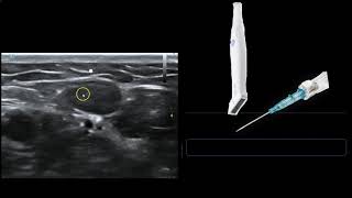
05:00 Demonstration of dynamic ultrasound-guided needle positioning on a simulator
06:51 Prevention of pathogen transmission via ultrasound probe
07:58 Ultrasound-guided needle repositioning
09:38 How to make a home-made simulator
10:59 Take-home Message
*************************************************
As MAGIC guideline suggests, with the aid of ultrasound-guided technique, short catheters and midline catheters should be used more often instead of PICCs and CVCs in the case of difficult vascular access.
The dynamic needle tip positioning technique can be applied to ultrasound-guided cannulation of virtually all peripheral vessels, including hemodialysis arteriovenous fistula, upper arm vein for PICC and midline catheter, and radial artery for monitoring. One exception is calcified arteries (e.g., radial artery of dialysis patients): In such cases, the “needle blinking method” may not work because the ultrasound beam does not penetrate the calcified wall.
A nephrologist in Kyoto, Tadashi Kamata, MD
************** Relevant RUclips video **************
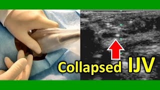
How to Safely Cannulate a Difficult Internal Jugular Vein on the First Attempt
• How to Safely Cannulat...
how to make & practice with an ultrasound phantom for IVs, lines, abscess
• how to make & practic...
Peripheral Vein Cannulation Using Ultrasound- transverse or longitudinal - which is best
• Peripheral Vein Cannul...
Ultrasound-Guided Peripheral IV Placement
• Ultrasound-Guided Peri...
Peripheral Venous Access Under Ultrasound Guidance - Part 1 - SonoSite, Inc.
• How to: Peripheral Ven...
ultrasound guided peripheral IV course by Siegfried Emme
• ultrasound guided per...
Ultrasound-guided arterial line Placement
• Ultrasound-guided arte...
PICC LINE INSERTION FULL PROCEDURE
• PICC LINE INSERTION FU...  Наука
Наука


This is probably the best ultrasound guided access content I've seen. I just wish you had some audio. Great work!
This essential ultrasound concept is not recognized by many other videos out there.
Thank you for your kind feedback. I would appreciate it if you could expand the method.
Dear Tadashi,
Thank you so much for this wonderful resource. Prior to this video, I was struggling to visualise the tip of my needle under ultrasound but now am able to guide the needle (the probe being perpendicular to the needle) the whole journey into the lumen of the vessel. Fan from New Zealand!
Hi, JYTS1970. Thank you for your kind comment. I'm very glad to be of service.
Great explanation. Thank you very much… 👍👏🙏🏿🙏🏻🙏🏻🙏🏻
what a wonderful instructional video !! I cannot thank you enough !! Hat off to you Tadashi--
Thank you for your kind feedback.
Very nice.Thanks.
Excellent video. Very good thank you
This is awesome
Thank you for this fantastic video
Thank you for your kind feedback.
Thank you !!! great video.
Thank you for your kind comment.
I always had a problem to visualize the needle tip on the screen, now I know I need to hole the probe tilting a little bit. A great video! Thanks!
Thank you for your kind comment.
Thank you a lot.
Thank you for your feedback.
excelent and intuitive information , thnakfully for share
Thank u sir
Picc Team here. Great video. Thank u.
Thank you for your comment. I'm glad you liked the video.
Nice video.
thanks very much, its very helpfull
Great! Thanks a lot!
Thank you for your kind comment.
very good video!
Thank you for your positive comment.
That was really useful - I will try the orthogonal method
Thank you for your kind comment. The point
of the orthogonal method is that using a sufficient amount of ultrasound jelly is essential to obtain a good image.
Thank you for this awesome video. I just got stumped after an annoying failure on a septic pt. Your guide will hopefully improve my technique for the future!
Thank you for your comment. I'm glad if my video is helpful.
Thanks
Thanks. How did you make the phantom?
The recipe is as follows, but unfortunately, it seems that cuboid konjak is only available in Japan.
1. To make mock vessels, build tunnels through a hard, cuboid konjac jelly by pressing a large straw into it.
2. Place the jelly in a tray whose depth is approximately the same as the thickness of the jelly.
3. Pour water into the tray until the top of the jelly is submerged.
4. Remove the air within the tunnels by squeezing the superior surface of the jelly with fingers.
😍🙏🙏🙏🙏😇
🇰🇭🗺️🌐
K
Where is the sound?
Sorry, no sound is available. I'll try someday.