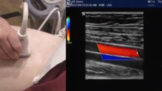
Liver Ultrasound: Using Shear Wave Elastography and Attenuation in diagnosing Fibrosis & Steatosis

HTML-код
- Опубликовано: 7 окт 2024
- Using the novel technology to diagnose liver fibrosis/cirrhosis & steatosis. This tutorial walks you through how to perform 2D SWE & Attenuation as well as how to position the probe for optimal viewing of the liver.
For sales inquiries please email sales@AdvisorMedTech.com
info@iSLUS.org
International Society of Liver Ultrasound


Good and easy explanation 👌
What is difference between elastography and fibroscan?
Hello, thank you for providing this video. This video, although I appreciate the presentation of it, seems more like a “how to do Elastography / SSP+ on a Hologic Ultrasound machine” rather than a general talk about SWE Elastography and using attenuation calculations to assess fatty liver infiltration. I also think it becomes an incredibly slippery slope if you are trying to differentiate F2 from F3 because Elastography data is not designed for intermediate differentiation like that, at least at this point in technological development/standardization. Perhaps renaming the video to “how do use a Hologic US Machine to perform SWE and SSP+” would reach an audience looking for this focused information instead. All vendors have different ways of acquiring this data. Thank you.
Thanks for the feedback Steven. I appreciate your comment since you work for Philips Ultrasound. We have reached out to your company for their involvement and welcome them to be apart of the discussion.