OPTIC DISC CHANGES IN GLAUCOMA
US
Войти
HOW TO READ AN OCT PRINTOUT IN GLAUCOMA || BASIC TESTING PROTOCOLS|| ( RNFL, ONH & MACULAR ANALYSIS}
23:38
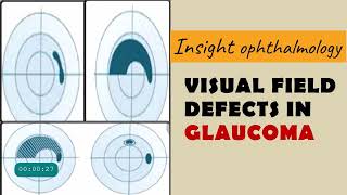
FIELD DEFECTS IN GLAUCOMA | arcuate, paracentral, nasal step, temporal wedge, baring of blindspot...
29:47
Eyes on the Future: Glaucoma Research Update from the 2024 Glaucoma Patient Summit
22:05
I.N "HALLUCINATION" | [Stray Kids : SKZ-PLAYER]
02:56
Every Form of Animation
24:43
Madison Police identify school shooter as 15-year-old female student
09:18
OPTIC DISC CHANGES IN GLAUCOMA
Insight Ophthalmology
Подписаться
59 тыс.
Скачать
Готовим ссылку...
Просмотров 47 тыс.
0
0
Добавить в
Мой плейлист
Посмотреть позже
Поделиться
Поделиться
HTML-код
Размер видео:
1280 X 720
853 X 480
640 X 360
Показать панель управления
Автовоспроизведение
Автоповтор
Опубликовано: 28 дек 2024
Комментарии • 57
Следующие
Автовоспроизведение
23:38
HOW TO READ AN OCT PRINTOUT IN GLAUCOMA || BASIC TESTING PROTOCOLS|| ( RNFL, ONH & MACULAR ANALYSIS}
Insight Ophthalmology
Просмотров 98 тыс.
29:47
FIELD DEFECTS IN GLAUCOMA | arcuate, paracentral, nasal step, temporal wedge, baring of blindspot...
Insight Ophthalmology
Просмотров 42 тыс.
22:05
Eyes on the Future: Glaucoma Research Update from the 2024 Glaucoma Patient Summit
Glaucoma Research Foundation Videos
Просмотров 6 тыс.
02:56
I.N "HALLUCINATION" | [Stray Kids : SKZ-PLAYER]
Stray Kids
Просмотров 1,9 млн
24:43
Every Form of Animation
TheOdd1sOut
Просмотров 3,7 млн
09:18
Madison Police identify school shooter as 15-year-old female student
Eyewitness News WTVO WQRF (MyStateline)
Просмотров 325 тыс.
34:13
MAKING BURR BASKETS FOR EACHOTHER!! ft: EVELYN ORTIZ
Blesiv
Просмотров 482 тыс.
1:06:54
High Yield Topic : Optic Disc Changes in Glaucoma - The Complete Course
Pranesh Balasubramaniam
Просмотров 103 тыс.
35:51
Lecture: Examining the Optic Nerve
Cybersight
Просмотров 11 тыс.
17:16
Vitamin B3 is the NEW Holy Grail of Glaucoma Treatment. Here’s Why.
Michael Chua, MD
Просмотров 131 тыс.
17:17
HYPERTENSIVE RETINOPATHY || pathology and signs
Insight Ophthalmology
Просмотров 25 тыс.
51:01
Retina | An Introduction | Part 1
Dr. Najeeb Lectures
Просмотров 839 тыс.
23:53
ANATOMY OF OPTIC NERVE
Insight Ophthalmology
Просмотров 28 тыс.
1:00:07
Prospects for Restoring Vision in Patients with Glaucoma: The Promise of Optic Nerve Regeneration
The Glaucoma Foundation
Просмотров 76 тыс.
32:02
9 EXAMINATION Optic Nerve Head and Nerve Fiber Layer Changes in Glaucoma
Dr.Mohammad Khabbaz
Просмотров 48 тыс.
59:38
Normal Tension Glaucoma: What You Need to Know (Webinar)
Glaucoma Research Foundation Videos
Просмотров 10 тыс.
20:38
СДЕЛАЛИ РЕМОНТ В ХИЖИНЕ ВОЗЛЕ НАШЕГО ПРУДА!
У Игоря
Просмотров 90 тыс.
1:20:25
BadComedian - Ненависть к VK, Блокировка YouTube, Что дальше? | Жубанион
ANOIR
Просмотров 791 тыс.
00:31
САЛЛИ УКРАЛА КРЕСТ! в отеле ДОРС роблокс | DOORS FLOOR 2 roblox | Секреты и приколы #Shorts
Red Cat
Просмотров 377 тыс.
00:59
СНЕЖИНКА (смешное видео, прикол, юмор, поржать, смех)
Натурал Альбертович
Просмотров 1,5 млн
2:09:03
Царица // 9 выпуск. Премьера
Телеканал ПЯТНИЦА
Просмотров 243 тыс.
25:48
Впервые Пробую ДУБАЙСКИЙ ШОКОЛАД и Другие ВИРУСНЫЕ СЛАДОСТИ 2024
Yan Reyzen
Просмотров 233 тыс.
00:22
CHOCOLATE MAGIC
MasomkaMagic
Просмотров 2,1 млн
00:33
Песня «Jingle Bells» на русском!🔔
Саша Квашеная
Просмотров 144 тыс.


![I.N "HALLUCINATION" | [Stray Kids : SKZ-PLAYER]](http://i.ytimg.com/vi/n5B5q1Hwt_U/mqdefault.jpg)