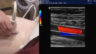
How to- Ultrasound duplex of the lower extremity arteries

HTML-код
- Опубликовано: 14 окт 2024
- This presentation provides basic ultrasound imaging of the leg arteries.
How to image and identify the lower extremity arteries using ultrasound,
Diagnostic criteria of arterial stenosis and how to identify a normal and abnormal arterial leg waveform
For more information visit www.divinescanning.com
My review book, Vascular Technology Made Simple is available on Amazon


Nice again. Fantastic video
Thank you
Excellent
Thank you
When you scan the pop where does your index on the probe point since your scanning from the posterior? Cus typically it’s always pointed to the patients right but does that apply when scanning from the posterior?
When scanning in a sagital view, the index points towards the head of the patient, when scanning transverse, the index regardless of what view you are utilizing points towards the sonographer(
Is the ATA flow always Biphasic ?
ATA flow is not always biphasic but can be considered normal with a normal ABI. There are 3 arteries feeding the foot. In a case were a biphasic ATA is seen and considered normal. PTA and Per are likely the dominant artery. Also certain heart conditions or scanning in a low temperature room can alter waveforms.
What’s some of your ways to find the ATA? I have trouble finding that. And also longitudinal SFA & SFV when it splits. I have trouble any tips please
I always locate my calf vessels from the ankle, for the ATA place your probe sagital on the tibia bone and angle laterally slowly you will run into it. I am guessing you meant the SFV/DFV, you can start trv with the CFV and slowly turn sag. the moment you see it split. The key is slow movements and less pressure when imaging your veins. A copy of my book will provide protocols and landmarks.
Please, why do choose to start the PTA in the distal region? Thanking you
It is sometimes easier to visualize in the smallest part of the limb, and also an abnormal waveform in the distal segment prompts the technologist to interrogate the PTA further
Hl. Dr. All. Vedlo