Femoral Triangle - Everything You Need To Know - Dr. Nabil Ebraheim

HTML-код
- Опубликовано: 31 июл 2018
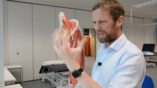
- Dr. Ebraheim’s educational animated video describes the anatomy associated with the popliteal fossa - posterior knee.
Follow me on twitter:
#!/DrEbraheim_UTMC
The femoral triangle is a superficial triangular space located on the anterior aspect of the thigh just inferior to the inguinal ligament. The boundaries of the femoral triangle include the lateral border, the medial border, and the base. The lateral border is formed by the medial border of the Sartorius m. The Sartorius muscle is a thin, small muscle going from lateral to medial. Sartorius muscle inserts on the medial side of the proximal tibia. The medial border is formed by the medial border of the adductor longus m. The adductor longus muscles goes from medial to lateral direction. The base is formed by the inguinal ligament. The floor of the triangle is formed by the iliacus muscle, the psoas major muscle, the pectineus muscle, and the adductor longus muscle. The roof of the femoral triangle is covered by skin, superficial and deep fascia. The femoral triangle contains three important structures (from lateral to medial) the femoral nerve, the femoral artery, and the femoral vein, and it contains the deep inguinal lymph nodes. NAVIgate the femoral triangle from lateral to medial. The femoral nerve lies within the groove between the iliacus and psoas major muscles. Two other nerves are located within the femoral triangle are the lateral cutaneous nerve of the thigh and the femoral branch of the genitofemoral nerve. The lateral cutaneous nerve of the thigh crosses the lateral corner of the triangle and supplies the skin on the lateral part of the thigh. The femoral branch of the genitofemoral nerve runs in the lateral compartment of the femoral sheath and supplies the majority of the skin over the femoral triangle. The femoral triangle also contains the femoral sheath which is a funnel shaped sleeve of fascia enclosing the upper 4 cm of the femoral vessels. The neurovascular bundle is medial to the Sartorius muscle. Therefore, in the anterior approach to the hip, it is always safe to go lateral to the Sartorius muscle in order to avoid the important structures within the femoral triangle. Do not go medial to the Sartorius muscle. You will injure the structures if you go medial to the Sartorius muscle. It is important to remember when performing this approach to avoid the lateral cutaneous nerve of the thigh. The Sartorius muscle is almost like the teres minor muscle in the shoulder. Do not go inferior to the teres minor muscle in the shoulder, you will injure the axillary nerve and the posterior circumflex artery.


Excellent information! 😉☝️ I'll have to put a video together on something similar soon! Thanks doc! 🤓👌
thanks for your efforts
I love your presentations . best wishs
i love you uncle ebraheim!
Be Fearless Hahaha
VERY INFORMATIVE BEST CHANNEL
Nice one....u r the stright way for medicin perpose.....i requst u to make for varicose in testis and leg....my humble request
best wishs