Cross sectional and imaging anatomy of the abdomen

HTML-код
- Опубликовано: 4 окт 2024
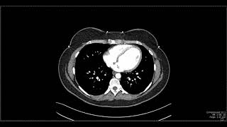
- This video deals with the anatomy of abdominal viscera and walls as they appear in transverse anatomical sections and axial CT sections. The video begins with a transverse anatomical section at the level of the T8 vertebra and continues down to the level of the L5 vertebra. There is a special emphasis on sections at the level of T10, T12, L1, L3, & L4.
00:04 Introduction
01:00 Section at the level of T8 vertebra
4:05 T10
8:30 T11/T12
12:45 T12
15:38 T12/L1
30:19 L1
32:29 L1/L2
36:25 L2/L3
39:35 L3
44:01 L4
The anatomical sections are arranged to match CT & MRI sections to provide a better understanding of the imaging anatomy of the abdomen. The arrangement of abdominal structures is followed in (22) serial transverse sections of the abdomen and compared to 5 representative axial CT sections at different levels.
The anatomical sections are selected from the Visible Human Project. For more information about this project refer to: www.nlm.nih.gov...
Five short video clips are inserted where necessary to explain the 3-D relations necessary to understand the 2-D sections.
Presented and edited by Dr. Akram Jaffar (Ph.D.). Plastic model clips filmed by Parwiz Akbari (medical student). Filmed at the College of Medicine/ University of Sharjah, UAE. 2012.
This video and its channel are supported by the "Human Anatomy Education" page on Facebook / anatomyeducation
Related videos:
Cross-sectional anatomy of the female pelvis and perineum • Cross sectional anatom...
Cross-sectional and imaging anatomy of the thorax • Cross sectional and im...
After completion of this video session, it is expected that you will be able to identify the approximate vertebral levels of transverse sections from T8-L5 vertebrae. The following structures are identified in transverse anatomical sections or CT axial sections:
Vessels:
Inferior vena cava (IVC), aorta, esophagus, azygos vein, hepatic vein, portal vein, hepatic artery, splenic artery, splenic vein, celiac trunk, renal vein, superior mesenteric artery, superior mesenteric vein, common iliac artery, basivertebral vein.
Viscera:
Lungs, liver, spleen, gastroesophageal junction, fissure for ligamentum venosum, gallbladder, caudate lobe, porta hepatis, common hepatic duct, cystic duct; pancreas: head, neck, uncinate process, body, and tail; spleen, hepatic flexure, splenic flexure, kidney; duodenum: first, second, and third part; stomach, pyloric antrum, pyloric canal, pyloric sphincter, ascending colon, transverse colon, descending colon, cecum, jejunum, ileum, plicae circularis, rugae, umbilicus.
Musculoskeletal structure:
Costal origin of the diaphragm, crura of the diaphragm, rectus abdominis, external oblique, internal oblique, transverse abdominis, linea alba, serratus anterior, rectus sheath, latissimus dorsi, trapezius, erector spinae, posas major, quadrates lumborum, pectoralis major, thoracolumbar fascia, ilacus, gluteus medius, spinal cord, cauda equine, filum terminale, costovertebral joint, costotransverse joint, ribs, costal cartilages, intervertebral disc, sternum, iliac crest.
Other related videos
Coronal section 1 • sectional anatomy of t...
Coronal section 2 • sectional anatomy of t...
Coronal section 3 • Sectional anatomy of t...
Coronal section 4 • Sectional anatomy of t...
sagittal sections
• Sectional anatomy of t...
Related accounts
Twitter / akramjaffar
Facebook / anatomyeducation
SlideShare www.slideshare....
LinkedIn / akram-abo. .
Research gate www.researchga....
Medtube medtube.net/us...
Instagram / akramjaffar
Academia dal.academia.e...


TO be honest, this is thebest teaching material ever produced to clarify understanding of CT scanning.
Thank you very much and God bless you.
As a medical student I can say this is one of the best made videos on medical imaging I´ve seen. Very helpful. Well worth a watch or two.
I'm not a Doctor, student or Radiologist, but this helped me understand ( To a certain extent ) my own CT scan, thanks a lot for posting such useful information.
I am a surgeon from airoli navi mumbai in India .
I wd like to congratulate you for an excellent presentation .
I am thankful and grateful to you for sharing this info ..It has really helped me conceptualise and read CT and MRI images .
Thank you and God bless you
+dr hemlata doke Thank you and best wishes.
The best demonstration of anatomy of abdomen
COMPERATIVE DEMONSTRATES OF CAT SCAN AND MRI
So nice of you, Thanks!
4 years later still benefiting from this video. Sincerely, Thank you !
very informative and clear, I wish all doctors explained as clearly as you did
Feel like i learn more from his videos than i do in class LOL. Thanks for making it easy to understand
Best wishes
yep i used this video too :)
Yeah... so why do people pay so much for school....... .... .....
This is one of the most helpful cross-section anatomy videos I've come across and I can't thank you enough for sharing! I am currently prepping for the CT registry and before I stumbled across your videos, I was feeling very nervous about the test and now I know I'll be okay. Thank you again!
Glad it was helpful!
I really appreciate this and how everything is executed.
I'm studying MRI in the fall and I suspect these videos will be invaluable.
Best wishes.
Absolutely stunning demonstration of human anatomy. Congratulations Dr. Akram for your efforts; I'm in fact relearning anatomy.
+Ashrarur Rahman Mitul Thank you for the compliments!
Sir, thank you for making this wonderful video.. Really helpful for aspiring radiologists and Radiologic Technologists !!
2:27am ⏰ , I feel like watching the video all over again. I enjoyed your teaching. Thank you ☺️
Glad you enjoyed it!
How I envy modern medical students who have the benefit of such superb anatomic learning tools. They will never know the countless hours spent learning the details of human anatomy that are so much better and more efficiently displayed now.
Proud to be one of your former students back in 1997 in Baghdad. God bless you❤🙏🏻
Thank you. All the best wishes!
Fantastic video, excellent for a medical student trying to get a basic idea of the normal anatomy for correlation with radiology!! Thank you so much : )
THANK YOU SO MUCH ! THIS HELP ME OUT SO MUCH FOR MY CROSS SECTIONAL CLASS.
Thank You, Sir! You are saving my sanity!!!! Wish You were my teacher!
Thank you for making such a great teaching video!! Best in youtube for CT anatomy.
Can't thank you enough for the wonderful videos you have created, it's so much easier to learn and remember the anatomical details and correlating it visually with Radiological approach is more easier, thank you very much sir, and your whole team
One of the ever Best Video about demonstrating CT abdomen...
HIGHLY recommended..
Would be more awesome if Pelvic anatomy was too demostrated...
I reckon you are one of the best Anatomist...
proud of you.
respect from Pakistan
I have plans to cover the male and female pelvic anatomy. Thank you for encouraging me to do so.
Very pratical video from anatomy to CT imaging.
+JL Tang best wishes
Thank you a lot Dr akram, one of the most instructive vidéo imaging learning i have seeing
So nice of you
What a presentation.... best presentation ever seen... may Allah bless you
+Ali Hossain Thanks!
This is a Useful video
I currently watched it now.
It helped me a lot in my CT-SCAN subject
Ang Hopefully i can passed the PRACTICAL EXAMINATION with 20 stations, and each of it contains
5 different structures that i need to identify.
God bless me ❤
Good Luck!
ppl like u r sunshine to the world....
keep goin....
Really very nice resource I hope it help me for my cross sectional anatomy for next summer Thank you so much for metrication prevention !
Many thanks from Singaporean medical students!
thank you very much sir.for making things very easy to understand
perfect anatomy teaching !!!!
wish you the best DR Akram
Thank you Ali. All the best for you too.
very nice presentation thanks alot ,Feel like i learn more from This videos.Thanks for making it easy to understand
you're a super medical teacher, thank you...
Thank you for taking the time out to make these helpful study tools : )
jazakAllah,very intresting and helpful
A big Thanks from India❤
You're welcome!
Seeing the body in cross section like this was eye opening even for an old doctor like me.
This is just so well explained. Thank you!
Glad it was helpful!
nice work, you did a so amazing dual-explain effort, much help from you, thanks a lot
There is enough evidence to show that it is the level of L3 like for example the presence of the right crus of the diaphragm and absence of the left crus, the presence of the third (horizontal) part of the duodenum etc.. However, lumbar vertebrae are quite thick (about 3cm) in addition to the intervertebral discs, thus the levels are not that sharp. Thank you for your comment.
Thank you so much for this Dr. Jaffar!!! It helped so much!!!!
ليتني بس عارفه عن الفيديو قبل فترة لأنه جدا جدا مفيد وفهمت فيه أشياء كنت مستصعبتها قبل :(
شكرا يا دكتور ، اشتراك +
what a great and useful video THANK YOU!
You can clearly describe the anatomy and thanks you so much
So helpful! Your a wonderful teacher thank you!
Thank you for this incredible educational video
6 years later am here, and im just grateful for your DR, thank you
asking for pelvis cross sections if available already
thank you and bless
Thank you! I have this project in mind and have the anatomical sections for both male and female bodies but not radiographs.
thank you soo much doctor that was very helpful ...we are waiting for more of your great videos :)
Many Thanks. Couldn't have been any better...Jazak Allah...
+ARSHAD HAYAT good luck!
very nice...well done doc!
Really it is the best. thank you sir for your effort
Thanks! you are welcome
thank you doctor it is very helpful video
awesome presentation, thankfull and greatfull to u sir
Thanks a looooooot sir!!!!it helped me a lot!!!!!your vid is a true guide for beginneers like me..wish to see more n more vids from you..immense regards from india... :)
this is amazing. thank you so much sir.
Thank you very much!
You're welcome!
This is mesmerising video NYC work Sir Allah bless u 🎉🎉
+Angel Libra thank you
amazingly described thank u soo much it was really helpful
Thank you!
ruclips.net/channel/UC_jGNnK94Pbfp-LRK5w_diAcommunity?lb=Ugwx1uo_bIJfYZHaT-V4AaABCQ
awesome! Tyvm! God bless you
What a master!!! thank you very very much
thank you so very much..your teaching is amazing!!!!!!!!!!!!!
Really great .Thank you so much for this very informative and clear explanation.Yet i am really astonished from the 45 thumb down persons !!! What do they need more clear and wide explanation better than this ? They are either ignorants or jealous .
So nice of you
THANK YOU SO MUCH SIR... EXCELLENT EXPLANATION..
u r great. I have seen some of ur other videos and they r all very very helpful
There ya go your 1000th like.... I have a mri of the abdomen in 2 days and as with other scans like to learn just enough to scare myself.
Dr. Akram Jaffar, your two videos of cross sectional and imaging anatomy are amazing! I've learned a lot from them. I was wondering if you have videos of cross sectional and imaging anatomy of the pelvis? Or Head & Neck? Thanks again
I am planning to do so. Subscribe to the channel and stay tuned.
Totally absorbed in it!
First class work.
Amazing, Amazing and amazing again. Please continue cause you convert sectional anatomy to a piece of cake for me and surely for many others. I am still confused that how we can have all those structures at the left side of the reverse. Can you explain it more in the comments? Its an axial section but I tried to look at it from the top view of the reverse and cannot understand why is that for? why we have the images in those forms?
When you look at CT or MRI axial images, you're viewing them as if you're looking up from the patient's feet towards their head. Therefore, the right side of the image corresponds to the patient's left side, and the left side of the image corresponds to the patient's right side.
The tradition for this orientation has its roots in older imaging modalities, like plain film radiography, where chest X-rays, for example, were placed on lightboxes to simulate the patient standing in front of you. This way of viewing images became standardized in radiology and was subsequently adopted for cross-sectional imaging such as CT and MRI.
For clinicians outside of radiology, this might be counterintuitive initially. It's important to always pay attention to any labels on the image that indicate "right" or "left" to avoid confusion when interpreting the scans.
amazing as always
Thank you!
Amazing work Doctor, i was waiting for it all this time :)
Great video! Thanks a lot.
Very good.
very nice presentation
عاشت ايدك دكتور اكرم
great video please upload more
Thank you sir !! It's exactly what i was looking for !!
FANTASTIC. Thank you!
Excellent video!
+jason marchione thanks
thank you so much Doctor!!!
Brilliant job 👍
Thank you! Cheers!
Excellent
very nice presentation thanks alot
FANTASTIC VIDEO!!!
Glad you liked it!
@@akram.jaffar Seriously. It made everything fall into place for me in imaging. Can you do a coronal section?
Coronal sections
Coronal section 1 ruclips.net/video/tiSDbUp7QbE/видео.html
Coronal section 2 ruclips.net/video/1oCyEtDarx8/видео.html
Coronal section 3 ruclips.net/video/1STRGIRxVOw/видео.html
Coronal section 4 ruclips.net/video/Dy0N8ZK52_8/видео.html
sagittal sections
ruclips.net/video/rL1SLcxRwBo/видео.html
Thanks for the explanation
You're welcome
you are too good sir, thank you
This was amazingly explained, thank you so much!! I was just wondering isn't esophagus usually flat in cross section why isn't it in these scans?
Good question! Check out this video ruclips.net/video/w-hhQNXQawU/видео.html to see how such cadaver was specially prepared. Please note that the video refers to the female cadaver and not to the cadaver used in imaging video which belongs to Joseph Paul Jernigan who donated his body after execution.
Excellent video. Cordial thanks sir. I would request you to cover other parts of the body like pelvis, Head, Neck & limbs.
+Dr. Md. Shalah Uddin will be done. Thank you.
thank you for this effort
Great thank for sharing knowledge
That was fascinating
Thanks!
Excellent!!!!
Many thanks!
Very fascinating video. Fantastic job. Dr. Jaffar i have a question regarding the thickness of the the space (fat) between a) the 2nd part of the duodenum to the anterior surface of the right renal vein and b) the space between Jujenum and the anterior surface of the left renal vein. Approximate numbers if possible. Thanks..
Wow, this was fantastic!
Did you happen to make a video of the pelvis or extremities? I can't seem to find them. These videos are very helpful!
No; but there is one on the thorax ruclips.net/video/9ftD8nLLOw8/видео.html
So helpful!! Thank you so much!!
thank you so much
+Vinay Reddy :-)
THANK YOU SO MUCH SIR....
You are welcome!
Thanks for sharing
My pleasure
Thanx Dr , amazing job
dr akram pls make a detailed video on the transverse and oblique pericardial sinus.It is really confusing...God bless u