Fetal Echocardiography - Exploring the Heart of the Unborn

HTML-код
- Опубликовано: 23 сен 2024
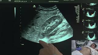
- Fetal echocardiography is a way to check your baby heart when you are pregnant. It helps see how
the heart is formed and working. It's done through a belly wand or a vaginal probe between 18 and
24 weeks.
Doctors recommend it when:
A standard ultrasound suggests heart issues.
There's a family history of heart problems.
You take certain medications, use alcohol or drugs.
You have conditions like rubella or type 1 diabetes.
How it's done:
The process is much like a regular ultrasound. A gel helps the wand pass sound waves through your
belly. These waves bounce off your baby's heart, and the wand picks them up. This creates detailed
pictures showing how the heart moves and how the blood flows. The test can also be done through
the vagina. It might take around 30 minutes to 2 hours.
Risks:
There are no known risks because it uses safe ultrasound waves.
#fetalecho #fetalechocardiography #echocardiagram #fetalechocardiogram #ultrasound


Excelent💝
Piniyal disorder explain karen
Awesome video 😊
Dear sactica par video Bana den
I love Biology 🧫 💕 So I watch your videos daily❤
Subhanallah
New information interesting ❤
Wa alikum assalam, Brother
👍
M 1st viewer 🐣🐣🐣
❤
Bhaijaan muje animation course karna AAP plzz suggest kare muje ❤
👍
👍