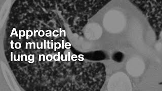
Honeycombing are dilated air spaces at the lung periphery and usually lung bases. Usually they are small and the walls between honeycombing are thick (fibrosis). Paraseptal emphysema will have thinner walls and usually are in the upper lungs. Subpleural cysts are usually much fewer in number than honeycombing. Emphysema and honeycombing can co-exist in the same patient too.
Thank you for your good work. You do a great job of explaining things that make it easy to understand and remember. Sometimes I find myself struggling to differentiate between honeycombing from UIP and subpleural cystic regions from severe emphysema. Any words of wisdom for telling the difference?
Yes, it can be very difficult some times esp since we know that a lot of patients with IPF are also smokers. In general, the craniocaudal location is helpful. Upper lung goes more with emphysema and lower lung goes more with honeycombing. Also, with honeycombing, the little septations between the dilated air spaces are usually thicker than what you see with emphysema. Emphysema, it is very thin wisps of tissue versus honeycombing which is thicker. Also, honeycombing tends to be smaller spaces compared to paraseptal emphysema in my experience.
Question. pleural plaques are common in asbestosis. Sarcoidosis is usualy mid lung perilymphatic - both are in alternative diagnosis yet you say that both diagnoses are typical UIP. Same goes to hypersensitivity pneumonitis which pattern is also differen to UIP. I dont understand how these are in typical UIP category 10:30
Good question. These entities have a range of appearances. The classic appearance won't be confused for UIP, but not every case has the classic features. Also, most cases of radiologic UIP will have a clinical dx of IPF, but those things are in the ddx.
Excelente video. Les dejo para complementar una lista de enfermedades que pueden causar Neumonía Intersticial Usual que creo que puede serles util. Saludos !! ruclips.net/video/PqQI-8Ztei0/видео.html


Excellent,crisp and very informative talk,like your voice .Thanks
Please we want more videos. Your teaching is excellent. Thanks for your good works
Excellent teaching .Thanks a ton
Great video!! Excited for the ILD series
Thanks for watching!
Many thanks for your excellent and useful videos. 🙏🙏🙏🙏🙏
thanks Sir, very good lecture, easy to understand.
This is Gold. 👏
excelent video regards from panama... this is a must see video for my residents
Excellent, great job sir
Thanks a bunch, was very didactic and well exposed
Great video. Beautifully explained. Many thnaks.
Nicely explained video..
Thanks for such a nice video sir
Hey, its really nice and crisp..
And Can u please elaborate on fibrotic NSIP vs probable UIP pattern?
Excellent! Thank you
You are welcome!
great! please do post more videos as they are exteremely useful
Master class
thanks. Waiting for more ILD
Beautiful ❤
Thank you Dr
Love your videos
Nice presentation
Can you suggest me a book to learn more about this topic ?
Excellent video !!
thanks. I like your voice
Please define honey-combing and how to differentiate with para septal emphysema and subpleural cysts.
Honeycombing are dilated air spaces at the lung periphery and usually lung bases. Usually they are small and the walls between honeycombing are thick (fibrosis). Paraseptal emphysema will have thinner walls and usually are in the upper lungs. Subpleural cysts are usually much fewer in number than honeycombing. Emphysema and honeycombing can co-exist in the same patient too.
@@ThoracicRadiology does honey combing have any exact definition ?? In terms of size, number , stacking ??? Thank you for your response!! Love it
Do you see cavitation with uip? Or Lip?
Thank you for your good work. You do a great job of explaining things that make it easy to understand and remember.
Sometimes I find myself struggling to differentiate between honeycombing from UIP and subpleural cystic regions from severe emphysema. Any words of wisdom for telling the difference?
Yes, it can be very difficult some times esp since we know that a lot of patients with IPF are also smokers. In general, the craniocaudal location is helpful. Upper lung goes more with emphysema and lower lung goes more with honeycombing. Also, with honeycombing, the little septations between the dilated air spaces are usually thicker than what you see with emphysema. Emphysema, it is very thin wisps of tissue versus honeycombing which is thicker. Also, honeycombing tends to be smaller spaces compared to paraseptal emphysema in my experience.
@@ThoracicRadiology thank you
Question. pleural plaques are common in asbestosis. Sarcoidosis is usualy mid lung perilymphatic - both are in alternative diagnosis yet you say that both diagnoses are typical UIP. Same goes to hypersensitivity pneumonitis which pattern is also differen to UIP. I dont understand how these are in typical UIP category 10:30
Good question. These entities have a range of appearances. The classic appearance won't be confused for UIP, but not every case has the classic features. Also, most cases of radiologic UIP will have a clinical dx of IPF, but those things are in the ddx.
Nice
hypersensitivity pneumonitis
Treatment start methylprednisolone 8 mg 2 tablet morning 15 days after 15 days 1 and half tablet 15 days....
Fvc 2.64. 73%
Fev1 2.22. 79 %
Feb1/fvc. 84.09
Female
height 160 cm
Weight 56
Age. 63
Please reply 100 % recovery possible?
Fvc 73. To. 80+ possible?
hypersensitivity pneumonitis cure possible?
hi, I'm not really sure. I think this is something you should ask your doctor.
Ophelia Mountains
The first video is the same as the second video
Karolann Bridge
Buford Key
75442 Micah Unions
Huels Haven
Mark Run
Carolyne Skyway
994 Isabell Walks
Carlie Drives
Smith Mews
Greenholt Mountains
Zackery Shore
Excelente video. Les dejo para complementar una lista de enfermedades que pueden causar Neumonía Intersticial Usual que creo que puede serles util. Saludos !! ruclips.net/video/PqQI-8Ztei0/видео.html
Baby Pike