Great video! IVC US is very useful in pts with spontaneous breathing but it has many limitations especially in mechanical ventilated pts then apart from focusing on physical exam we should not rely solely on IVC US but should combine it with lung US, cardiac US and internal jugular vein US.
Very well done! I often compare POCUS to driving stick shift. Subtle movements are the difference between driving smoothly and stalling. Your advice is well delivered. Perhaps it would be helpful to do an addendum with a curvilinear probe? From my experience it is easier to to use than the phased array or convex probe and can be an adjunct in obtaining images in patients who are very challenging. Also could you perform an example of passive leg raise to evaluate for fluid responsiveness?
Wonderful video! I read about using the motion mode to calculate the CI. Can you clarify that for me please? How we use M mode to calculate IVC collapsibility?
If you insert a CV line and measure the CV pressure directly they tell you it is unreliable, then they put a probe on the abdomen to estimate the CV pressure


This is by far the most useful YT channel for bedside clinical exams.
Thank you :-)
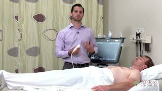
Dr. Kumar is an excellent patient.
Excellent demonstration Dr Kugler
Great video! IVC US is very useful in pts with spontaneous breathing but it has many limitations especially in mechanical ventilated pts then apart from focusing on physical exam we should not rely solely on IVC US but should combine it with lung US, cardiac US and internal jugular vein US.
Yes agreed! We make sure to cover all those in other videos. Thanks.
Wonderful. There is no a better feeling of that of learning something❤
Awesome video. Stanford putting up to its huge tradition in Medicine!♥️
Very well done! I often compare POCUS to driving stick shift. Subtle movements are the difference between driving smoothly and stalling. Your advice is well delivered. Perhaps it would be helpful to do an addendum with a curvilinear probe? From my experience it is easier to to use than the phased array or convex probe and can be an adjunct in obtaining images in patients who are very challenging. Also could you perform an example of passive leg raise to evaluate for fluid responsiveness?
Great ideas! Plan is to add more short videos to supplement these. Thank you.
Great video, thanks!
Great explain thanks Doctor
So good
Wonderful video! I read about using the motion mode to calculate the CI. Can you clarify that for me please? How we use M mode to calculate IVC collapsibility?
thanks for great explain 💯
If you insert a CV line and measure the CV pressure directly they tell you it is unreliable, then they put a probe on the abdomen to estimate the CV pressure
My thought exactly 😅
Is the hepatic vein the same one when measuring the diameter of IVC in the mid axillary line ?
What is the normal diameter? You said up to 2.5 is normal. I read in other places it’s 1.7
Great
The measurement is completely diagonal to the vessel, overestimating diameter by a lot. Otherwise it's a great instructional video.
💚
¿Alguien conoce una página de esta calidad pero en español?
Excellent video, but I need the volunteer guy to contact me, lol. Super cute!
Roob Court
Wrong way to measure diameter .caliper should be perpendicular to two side of IVC walls
Martinez Ruth Hall Steven Martin Ruth
Smith Ruth Taylor Carol Thompson Frank
Brown George Brown Nancy Lee Nancy