How To: Gallbladder Ultrasound Part 1 - Introduction Case Study Video

HTML-код
- Опубликовано: 24 июл 2024
- Go to www.sonosite.com/education for more videos and information about ultrasound technology.
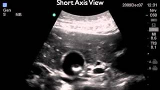
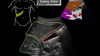
This video (part 1 of 3) details how bedside medical ultrasound imaging of the gallbladder allows for rapid evaluation of patients presenting with acute abdominal pain. The video focuses on normal hepatobiliary imaging, as well as the anatomy of and near the gallbladder.
To receive notifications about RUclips videos from Sonosite, click the Subscribe button to subscribe to this channel.
For a more comprehensive and portable video library, please download the SonoAccess™ iPhone® app at www.sonoaccess.com.
------------------------------------------------------------------------------------------
We recognize the value of point-of-care ultrasound better than anyone else, because it's all we do. FUJIFILM Sonosite is an innovator and leader in point-of-care ultrasound and our portable, compact systems expand the use of ultrasound across the clinical spectrum by bringing high-performance ultrasound to the point of patient care.
Sonosite LX: www.sonosite.com/products/son...
Sonosite PX: www.sonosite.com/products/son...
All Products: www.sonosite.com/products/ult...
Educational Resources:
Webinars: secure.sonosite.com/behind-th...
Sonosite Institute: www.sonosite.com/education/so...
Our Website: www.sonosite.com/
Social Media Channels:
/ sonosite
/ sonosite
/ fujifilm_sonosite
/ sonosite
#ultrasound #ultrasoundmachine #ultrasonography #portableultrasound #pointofcareultrasound #bedsideultrasound #ultrasoundtraining #clinicalsupport #ultrasoundvideo #ultrasoundguidance #ultrasoundeducation #ultrasoundcourses #ultrasoundapplications #medicalultrasound  Наука
Наука


As an American who now lives in the Philippines, imagine my 3 month ordeal being in the hospital where I had to teach the Sono-Technican, nurses, the language and terminology. My so called (wrongly diagnosed) Pancreatic cancer was actually a gallstone. I still have my bladder and have been symptom free for 3 years now after I escaped the hospital and used food to cure myself. And they wonder why even the locals travel to the USA for medical treatment. Excellent video !
ha ha ha....what happened to the gallstone?
It was reduced in size by lemon juice, Apple Cider vinegar, an a change in diet, then passed out into the intestines where it was expelled in a bowel movement. By the way, today is Jan 2014...no problems.
Lemon Juice and olive oil for the flush...but starting a week before everyday a shot of ACV in the morning... yes documented just google it home remedy liver flush sometimes sites vary in instructions..the Chinese way is the best in my opinion
RandomActUpMedia 8
RandomActUpMedia How are you now??
Fantastic! very helpful for the medical student
Excellent Reliable videos
great work sir
Excellent teaching.
very helpful
Thank you
hi! this might be a stupid question, but do you still measure the gallbladder WALL if the patient didnt fast and ate something prior to exam?
excellent
Thank you sir
My exam look nothing like this. It took them a few minutes to find it and then to force the little string of five or six pearls into the shot with nothing else in the sack and they formed a line and were perfectly round. I wonder if it is too early to have this organ just cut out? I can find a shot that looks anyting lime mine.
can you please do a RUQ ultrasound with liver measurement
Good presentation
great teaching
Awesome 5***** thank. You
had an ultrasound today to check my liver and gAllbladder as have been having pain under right rib and in right side of stomach since April when i was admitted to hospital with severe stomach pains.my daughter was with me at my scan and said there was a large red blob in a big black blob(she's only 10 lol) the lady also scanned my kidneys.i have to call Dr for results on Friday.if i have gallstones i will have my gallbladder removed.if its all clear then im still not sure what is causing my pain
Very nice 👍
@4:50 for example, the pic is clear enough, but the location and orientation of the probe that created that ultrasound image is not shown: very incomplete.
pregunto el modelo tittan de sono site esta descontinuado
Thanks
v nice
nice demo.useful for student sonologists
good
Thanks sir very very your vadio very help ful
Good
Mere bhi gallblader me 2 stone the 5mm and 8mm ke bohot hi preshan thi mai operation krne ki bilkul capicity nhi thi mere husbend ne youtub se ayurvedic khadnol Livcon capsule and khadnol syrap k bare me dekha to unhone amazone se mngvakr dr, se suggestion leke muze diya or isse mere stones 5 se 6 month me hi nikal gaye or tbbiyat bhi thik hai meri,,
i have undergo gall stone surgery . After completion surgery doctor show me gall stones of black colour.
tq
where are the sources from?
cool
im having the same problem. what happened? im getting my ultra sound on Friday
🔥
Good morning
Enjoyable
i wish it had been a female depiction....but very informative none of the less, well done and thank you for uploading.
Why female
Do they differ between sexes?
gallbladder cholestrol crystals ultrasound
Aorta ultrasound
Thanks sir