I got 8/10! I must say, I forgot to apply B rules on the ones where the simple descriptors were applicable. Looking forward to more videos. Thank you very much.
That's really great well done! There are more and newer videos on the prediction of malignancy in adnexal masses in this playlist ruclips.net/p/PLzdkYHOauxtS--51sqgVXbT0kaLPwjelF You are quite right, if the diagnosis is obvious on Simple Descriptors (eg dermoid or endometrioma) then you don't need to go to Simple Rules, or even ADNEX or O-RADS. You already have the answer :) But I often show all the different methods (of deciding if the mass is benign or malignant) just for practice :) Enjoy the other videos, the website is slightly easier to navigate gynaecologyultrasound.com/ Best wishes
Thank you so much! Here is a slightly more recent lecture, and do look at the website for more talks on other gynae ultrasound subjects. Best wishes gynaecologyultrasound.com/iota-inc-adnex/
Glad you liked it :) There are also newer videos on ‘How to predict malignancy in adnexal masses’ like this one ruclips.net/video/FrVpDQFMsRk/видео.html. Best wishes
Thank you so much :) There are lots more videos on the website www.GynaecologyUltrasound.com. Including more on IOTA (or on the YT playlist - I’ll send you the link :) Best wishes
Thank you so much - that was quite an old video - here is a playlist of more recent adnexal pathology ultrasound videos : ruclips.net/p/PLzdkYHOauxtS--51sqgVXbT0kaLPwjelF
definitely a thought provoking lecture. very very useful. Loved the ' quiz ' at the end, although i didnt fare all correct at it, but it has given me a path to think and implement. a fantastic session.. Now, after long we audience can ask, how did i miss this site of urs.. and i like the passion with which u answer most of questions put up. Thank you very much. Looking forward for more of ur videos and quiz style..Pranams.. - Shriraam Ayyar, India
Thank you so much - here is a playlist of videos about IOTA, which includes a more recent video about the prediction of risk of malignancy in adnexal masses, including the ADNEX risk model ruclips.net/p/PLzdkYHOauxtS--51sqgVXbT0kaLPwjelF
Could you please tell me what the ADNEX Model is and how it differs from IOTA and the associated logarithmic regression models? Once again thank you for helping me to understand these topics.
Again - how on earth did I miss these questions? Sorry! By now you may have seen this video, which hopefully addresses your questions? ruclips.net/video/27uwcytplpo/видео.html Best wishes (and I will try to look for all comments in the future!)
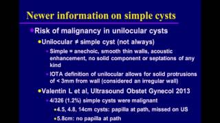
thanks for the great lecture one question please in 8:37 , what is described as subtle shadowing , couldn't me consider it is ( critical angle shadowing artefact ) rather than a real one ?
Thank you for your comments. During the scan it appeared as a real and fine shadow, rather than artefact. The main point is that one has to look for shadowing, that it can be dense or really fine (eg in cystadenofibromas) and that just because a mass has shadowing, doesn't make it benign (it is only a benign feature). Many cancers also have shadowing. There is more content on gynaecologyultrasound.com/ and also on Twitter twitter.com/GynaecologyUS Best wishes
@@magedalgazzar389 Using IOTA Simple Descriptors, an ovulatory corpus luteum will probably be instantly recognisable as physiological and be described as such. It is a unilocular cyst with no solid component but usually haemorrhagic contents with strong peripheral vascularity - although this strong vascularity ('ring of fire') can look alarming, the endometrium will also be in the luteal phase and this will guide you to the correct diagnosis. A CL does not need management or follow-up
@@GynaecologyUltrasound thanks for your kind help two questions please - in case of a corpus luteum (cyst) : the crenelated wall may be mistaken as soft tissue / wall irregularity in some cases can i recommend follow up - the descriptor of mature cystic teratoma : will it be enough to exclude an immature teratoma , i mean how can i differentiate the mature vs the immature teratoma via imaging thanks
@@magedalgazzar389 Sorry for the slow reply. Re corpus luteum - if you are uncertain about whether an appearance is physiological or not, you can always rescan in around 4 weeks and look for resolution/change. Re mature vs immature teratoma - generally speaking a mature teratoma(dermoid) will have typical features and low or absent vascularity, whereas an immature teratoma can have areas of strong vascularity and irregular solid components - it will look 'odd' at the very least. Also immature teratomas are quite rare and mature ones very common. Again, you could repeat the scan at an interval and look for change. If you have any doubt about the benignity of a lesion then always put that in the report so that the 'requesting clinician' can (potentially) request alternative imaging and management strategies.
First of all: thank you for this useful video. I have two question: - How could you determine (or what are the differences between) an uniloculair solid mass versus an uniloculair mass with a papillar structure? - How could you determine (or what are the differences between) a solid mass versus an uniloculair dark homogenous mass? Thank you in advance!
The definitions really matter. Any tissue that appears solid but is 3 mm is called 'Solid'. If the solid tissue indents the cyst (fluid on 3 sides) then it is a papillation. But if the solid component is flat then it is not a papillation. So all papillations are solid, but not all solid tissue is a papillation
And solid tissue may have vascularity on Doppler, but necrotic tissue doesn't. So if tissue is vascular it must be solid (rather than cystic) but not all solid tissue has vascularity. (If a cyst contains thick gloop which moves with the energy from the sound waves (called 'streaming') then that may show up as colour so be careful
So if a lesion has one locule and a solid component that indents the cyst (>3 mm height) then it is a unilocular-solid lesion (or you can say 'unilocular with solid')
it can be difficult to distinguish between a unilocular cyst and a solid mass. The cyst will let sound through so you get posterior acoustic enhancement (brighter behind) whereas the solid mass absorbs sound so you can get shadowing. But it can be difficult. If the lesion has strong central vascularity then it must be solid...
Hypothetically speaking, if you see what looks like a grossly normal dominant follicle of around 2cm but it has a single 4 mm papillation in a pre-menopausal woman, do you class it as normal? Similarly if you see cysts within cysts or follicles with daughter follicles within - are they classed as normal in pre-menopausal women?
Thank you. Be careful what you call a papillation - this is solid tissue of >3mm in height from the cyst wall. It has to be solid tissue, ie not clot (eg in a haemorrhagic cyst) or Rokitansky nodule (in a dermoid) or mucin (layered in a mucinous cystadenoma). Solid tissue has to have the appearance of tissue and may be vascular. What you describe sounds like a lesion within the ovary, and if the rest of the ovary is normal, you would describe this as unilocular-solid. The internal cyst wall is now irregular. You have not mentioned what the vascularity is like, or whether there is any shadowing. To apply Simple Rules, in the absence of any malignant features, such an appearance would count as a benign feature (solid material
No shadowing or vascularity. Thank you. I'm currently using IOTA for a uni assignment. There is a bit of a grey area in the adnexal lesion pathway at my study hospital in terms of what "non-simple lesions" should be referred for a gynae opinion and what should be purely repeated at interval. Obviously anything with malignant features would warrant a gynae opinion/CA125 in addition to initial ultrasound. Alternatively I've noticed practitioners may simply rescan a haemorrhagic cyst, or even not follow it up if it is deemed functional or under a certain measurement in a pre-menopausal patient. It's the benign lesions that I'm currently battling to get my head around ... oh and LR1 and LR2 now I've looked into some of the articles you recommended.Without a comprehensive understanding the rules I fear people could overlook inconclusive lesions as benign. As a result ... patients from e.g. GPs could slip through the net and not receive timely treatment. P.S. thank you for your explanation of solid material as non-solid materials could also be easily misinterpreted.
About FU of ovarian lesions - here are two excellent references for you: Management of asymptomatic ovarian and other adnexal cysts imaged at US Society of Radiologists in Ultrasound consensus conference statement. Levine et al , Ultrasound Q. 2010 Sep;26(3):121-31 and RCOG GreenTop Guideline 62 (2011) - Management of Suspected Ovarian ~Masses in Premenopausal Women and soon i will upload some protocols to the website www.gynaecologyultrasound.co.uk (being written at the moment) About Simple Rules - these can be used by sonographers when they have been agreed in your department and are understood by everyone - that is the reason I wrote these videos. I hope you have found them useful. After a scan in which there is diagnostic doubt, the advice to the referring doctor should be to 'refer to gynaecology'. Uncertain/inconclusive is definitely not the same as benign - where there is no 'expert opinion', if the mass is uncertain/inconclusive, then some centres treat all such cases as though they are malignant - that way you won't miss any/many malignancies (but will overcall some). good luck with your project


Amazing presentation mam.
Excellent presentation ,very much informative lecture ,thank you
Thank you very much. Excellent presentation, complex topic made simple & easy to understand & Remember.
I got 8/10! I must say, I forgot to apply B rules on the ones where the simple descriptors were applicable. Looking forward to more videos. Thank you very much.
That's really great well done! There are more and newer videos on the prediction of malignancy in adnexal masses in this playlist ruclips.net/p/PLzdkYHOauxtS--51sqgVXbT0kaLPwjelF
You are quite right, if the diagnosis is obvious on Simple Descriptors (eg dermoid or endometrioma) then you don't need to go to Simple Rules, or even ADNEX or O-RADS. You already have the answer :) But I often show all the different methods (of deciding if the mass is benign or malignant) just for practice :) Enjoy the other videos, the website is slightly easier to navigate gynaecologyultrasound.com/ Best wishes
Great 👍
Simple & excellent presentation !!! THANK you so much !!!
Thank you so much! Here is a slightly more recent lecture, and do look at the website for more talks on other gynae ultrasound subjects. Best wishes gynaecologyultrasound.com/iota-inc-adnex/
excellent vedio mam....very clear and informative. quiz part i loved it... thanks a lot
Thank you so much :) There are many more videos on the website gynaecologyultrasound.com/ Best wishes
Excellent lecture ,thank you
My sincere appreciation/gratitude/thanks🌹very helpful.
Glad you liked it :) There are also newer videos on ‘How to predict malignancy in adnexal masses’ like this one ruclips.net/video/FrVpDQFMsRk/видео.html. Best wishes
Thank you very much in deed for this very simplified explanation of iota
Thank you so much :) There are lots more videos on the website www.GynaecologyUltrasound.com. Including more on IOTA (or on the YT playlist - I’ll send you the link :) Best wishes
ruclips.net/p/PLzdkYHOauxtS--51sqgVXbT0kaLPwjelF.
Very helpful..thank you for sharing ..God bless
Thank you so much :). There are many more videos on GynaecologyUltrasound.com. Best wishes
Very nice, thank you for your time and effort 👍
Thank you so much - that was quite an old video - here is a playlist of more recent adnexal pathology ultrasound videos : ruclips.net/p/PLzdkYHOauxtS--51sqgVXbT0kaLPwjelF
Thanks a lot for this leacture! Very useful!
Thank you so much! There are lots more videos on the website - do have a look gynaecologyultrasound.com/
thanks for this wonderfull presentation
Glad you liked it. This is the most recent video in the prediction of malignancy in adnexal masses ruclips.net/video/vKhmKJZo23A/видео.html
definitely a thought provoking lecture. very very useful. Loved the ' quiz ' at the end, although i didnt fare all correct at it, but it has given me a path to think and implement. a fantastic session.. Now, after long we audience can ask, how did i miss this site of urs.. and i like the passion with which u answer most of questions put up. Thank you very much. Looking forward for more of ur videos and quiz style..Pranams.. - Shriraam Ayyar, India
Thank you so much - do spread the word! The linked website is GynaecologyUltrasound.com. Best wishes
Thanks . Great lecture!!!
F T thank you ! Do have a look at the website for more videos gynaecologyultrasound.com/.
Great video. Thanks to narrator
Manas Das You’re very welcome. There are more videos here : gynaecologyultrasound.com/
Good day ! 😢 my mother have a IOTA operation is answer of painful? Thank!
Hopely I get your reply
Judy!
Thank you Doctor. Very useful lecture
Thank you very much - do have a look at the website for more videos and information gynaecologyultrasound.com/
Very helpfull, thanks.
Thank you so much - here is a playlist of videos about IOTA, which includes a more recent video about the prediction of risk of malignancy in adnexal masses, including the ADNEX risk model ruclips.net/p/PLzdkYHOauxtS--51sqgVXbT0kaLPwjelF
Could you please tell me what the ADNEX Model is and how it differs from IOTA and the associated logarithmic regression models?
Once again thank you for helping me to understand these topics.
Again - how on earth did I miss these questions? Sorry! By now you may have seen this video, which hopefully addresses your questions? ruclips.net/video/27uwcytplpo/видео.html Best wishes (and I will try to look for all comments in the future!)
thanks 🎉
what's difference b/w serous & mucinous cystadenoma in usg? i can't got it 😢
HI - they can look very similar - have a look at this video to see the differences ruclips.net/p/PLzdkYHOauxtTRUoa_N1Oc5Ze1LKoX29xB Good luck :)
thanks for the great lecture
one question please
in 8:37 , what is described as subtle shadowing , couldn't me consider it is ( critical angle shadowing artefact ) rather than a real one ?
Thank you for your comments. During the scan it appeared as a real and fine shadow, rather than artefact. The main point is that one has to look for shadowing, that it can be dense or really fine (eg in cystadenofibromas) and that just because a mass has shadowing, doesn't make it benign (it is only a benign feature). Many cancers also have shadowing. There is more content on gynaecologyultrasound.com/ and also on Twitter twitter.com/GynaecologyUS Best wishes
Another question please ...how do u describe/manage a corpus luteum cyst using IOTA ?
@@magedalgazzar389 Using IOTA Simple Descriptors, an ovulatory corpus luteum will probably be instantly recognisable as physiological and be described as such. It is a unilocular cyst with no solid component but usually haemorrhagic contents with strong peripheral vascularity - although this strong vascularity ('ring of fire') can look alarming, the endometrium will also be in the luteal phase and this will guide you to the correct diagnosis. A CL does not need management or follow-up
@@GynaecologyUltrasound thanks for your kind help
two questions please
- in case of a corpus luteum (cyst) : the crenelated wall may be mistaken as soft tissue / wall irregularity in some cases can i recommend follow up
- the descriptor of mature cystic teratoma : will it be enough to exclude an immature teratoma , i mean how can i differentiate the mature vs the immature teratoma via imaging
thanks
@@magedalgazzar389 Sorry for the slow reply. Re corpus luteum - if you are uncertain about whether an appearance is physiological or not, you can always rescan in around 4 weeks and look for resolution/change.
Re mature vs immature teratoma - generally speaking a mature teratoma(dermoid) will have typical features and low or absent vascularity, whereas an immature teratoma can have areas of strong vascularity and irregular solid components - it will look 'odd' at the very least. Also immature teratomas are quite rare and mature ones very common. Again, you could repeat the scan at an interval and look for change. If you have any doubt about the benignity of a lesion then always put that in the report so that the 'requesting clinician' can (potentially) request alternative imaging and management strategies.
First of all: thank you for this useful video. I have two question:
- How could you determine (or what are the differences between) an uniloculair solid mass versus an uniloculair mass with a papillar structure?
- How could you determine (or what are the differences between) a solid mass versus an uniloculair dark homogenous mass?
Thank you in advance!
HI thanks for your lovely email :) There are more videos on this topic in this playlist: ruclips.net/p/PLzdkYHOauxtS--51sqgVXbT0kaLPwjelF
The definitions really matter. Any tissue that appears solid but is 3 mm is called 'Solid'.
If the solid tissue indents the cyst (fluid on 3 sides) then it is a papillation. But if the solid component is flat then it is not a papillation. So all papillations are solid, but not all solid tissue is a papillation
And solid tissue may have vascularity on Doppler, but necrotic tissue doesn't. So if tissue is vascular it must be solid (rather than cystic) but not all solid tissue has vascularity.
(If a cyst contains thick gloop which moves with the energy from the sound waves (called 'streaming') then that may show up as colour so be careful
So if a lesion has one locule and a solid component that indents the cyst (>3 mm height) then it is a unilocular-solid lesion (or you can say 'unilocular with solid')
it can be difficult to distinguish between a unilocular cyst and a solid mass. The cyst will let sound through so you get posterior acoustic enhancement (brighter behind) whereas the solid mass absorbs sound so you can get shadowing. But it can be difficult. If the lesion has strong central vascularity then it must be solid...
Thank you very much
Thank you so much - sorry I hadn't seen your comment. There are many more videos on the website gynaecologyultrasound.com/ Best wishes :)
Hypothetically speaking, if you see what looks like a grossly normal dominant follicle of around 2cm but it has a single 4 mm papillation in a pre-menopausal woman, do you class it as normal?
Similarly if you see cysts within cysts or follicles with daughter follicles within - are they classed as normal in pre-menopausal women?
Thank you. Be careful what you call a papillation - this is solid tissue of >3mm in height from the cyst wall. It has to be solid tissue, ie not clot (eg in a haemorrhagic cyst) or Rokitansky nodule (in a dermoid) or mucin (layered in a mucinous cystadenoma).
Solid tissue has to have the appearance of tissue and may be vascular.
What you describe sounds like a lesion within the ovary, and if the rest of the ovary is normal, you would describe this as unilocular-solid. The internal cyst wall is now irregular. You have not mentioned what the vascularity is like, or whether there is any shadowing.
To apply Simple Rules, in the absence of any malignant features, such an appearance would count as a benign feature (solid material
No shadowing or vascularity. Thank you. I'm currently using IOTA for a uni assignment. There is a bit of a grey area in the adnexal lesion pathway at my study hospital in terms of what "non-simple lesions" should be referred for a gynae opinion and what should be purely repeated at interval. Obviously anything with malignant features would warrant a gynae opinion/CA125 in addition to initial ultrasound. Alternatively I've noticed practitioners may simply rescan a haemorrhagic cyst, or even not follow it up if it is deemed functional or under a certain measurement in a pre-menopausal patient. It's the benign lesions that I'm currently battling to get my head around ... oh and LR1 and LR2 now I've looked into some of the articles you recommended.Without a comprehensive understanding the rules I fear people could overlook inconclusive lesions as benign. As a result ... patients from e.g. GPs could slip through the net and not receive timely treatment. P.S. thank you for your explanation of solid material as non-solid materials could also be easily misinterpreted.
About FU of ovarian lesions - here are two excellent references for you:
Management of asymptomatic ovarian and other adnexal cysts imaged at US Society of Radiologists in Ultrasound consensus conference statement.
Levine et al , Ultrasound Q. 2010 Sep;26(3):121-31
and
RCOG GreenTop Guideline 62 (2011) - Management of Suspected Ovarian ~Masses in Premenopausal Women
and
soon i will upload some protocols to the website www.gynaecologyultrasound.co.uk
(being written at the moment)
About Simple Rules - these can be used by sonographers when they have been agreed in your department and are understood by everyone - that is the reason I wrote these videos. I hope you have found them useful.
After a scan in which there is diagnostic doubt, the advice to the referring doctor should be to 'refer to gynaecology'.
Uncertain/inconclusive is definitely not the same as benign - where there is no 'expert opinion', if the mass is uncertain/inconclusive, then some centres treat all such cases as though they are malignant - that way you won't miss any/many malignancies (but will overcall some).
good luck with your project
Thanks a lot
I have my result today ive got B1 and M3 what does it mean ? is it malignant I 'm afraid
That means that Simple Rules is Indeterminate (uncertain) and you may need more tests. Best wishes
@@GynaecologyUltrasound thank you so much