Clinical Laboratory Engineering Specialist Florida Hospital Orlando, Florida. This was one of the best learning tools I have had the opportunity to view. I highly recommend it to Laboratory Technicians and service engineers alike. Well done. Russell McGahee ISA, CLES
Quick question for my general understanding? At around 03:00 you say that the greater the cell, the more light will fall on the detector and vice versa for smaller cells. My question is; Shouldn't it be the other way round, since the size of the detector won't change and the light will scatter more in other directions, thus leaving a larger 'shadow' behind on the detector? Would appreciate an answer and thus thank you in advance. However, thanks for the effort and the very nice demonstration on how this works! Stay motivated and curious! =)
Scattering appears not from the "borders" of the cell as it is in the pictures here, but from different intristic parts of the cell. So, if you have a larger cell, your detector will get scattered signal for longer time, thus the width of the forward signal peak (which is normally refered as FSC-W) wiil be greater than it is for a smaller cell. The height of the forward scattering peak (FSC-H, the maximum amount of light scattered during an event) could not be directly associated with sizes of the cells, but could be a marker which is unique for every type of cells. Normally we want to take in account both parameters (size and "type"), so we use their combination which is an area under the peaks curves (FSC-A).
So small molecules will give less FW scatter in comparison to large molecules? How does that work? I always thought it was the other way around; big molecules block more light then small molecules so give less FW scatter. Can u explain it to me pls?
The cells are so small to block the laser beam ... the small cells cause less forward scatter and the large cells cause more scatter (as shown in the video between 2:50 and 3:00) ...
It's less "block more light" and instead scatters the light. So as was said before, all of the cells are too small to block the laser beam entirely. But, instead, as they pass through, they change the angle of the light so that it's not going straight, but scattering it. But think of it like a water tap. If you pass a small thing through it (your finger, for example), the flow breaks and spreads out. But if you pass something much larger (your fist, for example), the same thing happens, but to a much larger area. So, a small cell will scatter/spread out the light as it passes through it, but a bigger cell makes a much larger area of light scatter as it passes through. Hopefully that made sense...
Usually the red blood cells are lysed and removed before running cells through the flow cytometer. In addition, we often set the threshold of a flow cytometer too high so that RBCs are excluded. If you were to keep the erythrocytes in the sample and set your threshold low enough to see them, they are very small and not granular, so they would show up at the very bottom left corner.
I think most important is that she explains everything very clearly and it's easy to understand and remember. There is a lot youtubers with better oral skills but their explanations isn't so easy to follow. So I prefer her "aaaaa" that some else's fluency
Such aberrated speech...can't focus at all...myself, being a stammerer, I re-record the broken parts of voice over as many times as required...and that is important!
You didn't do the best at explaining, a lot of "uhm"s and "uhhh"s, you were lost for your words half the time. In saying that the graphics is what saved this. I came here for a quick review so I knew what you were trying to say. I'd prepare a script for your next video.

 Кино
Кино


![The moment we stopped understanding AI [AlexNet]](/img/1.gif)
Your excellent explanation on how Flow Cytometry works helped me a lot today.
Thanks!
Clinical Laboratory Engineering Specialist Florida Hospital Orlando, Florida. This was one of the best learning tools I have had the opportunity to view. I highly recommend it to Laboratory Technicians and service engineers alike. Well done.
Russell McGahee ISA, CLES
Outstanding explanation of flow cytometry. Thank you!
I am a nursing stutend in my last year.
I enjoyed your's explenation a lot!!
Very clear and understandable. Thank you greatly for your lectures!
Thank you, for your powerful explanation that make everything easy to learn. Good luck🌹
ruclips.net/video/orBv5ZgsV8Q/видео.html
Very good job at explaining this procedure!
Thank for the simplicity of explanation
Thanks for the video on the flow cytometry. You are an amazing teacher
Thank you very much for your wonderful explanation that make anyone to understand Flow Cytometry. Good luck.
I really like to watch this video. I almost understand all the thing that you explained. thank you for this video!
Amazing..very helpful video.thank you
Very useful and didactic video. Congrats
Awesome explanation. Thanks
Thank you so much! This is very useful!!!
this made it very clear thank you!
Very well explained.. Thank u.
Great explanation, thanks !
Excellent explanation!! Thank you very much.
Excellent video. Thank you
Very clear and helpful! Thank you!
Thanks for your excellent explanation about flow cytometry
Very well and clear explained video... thank you ma
Very well explained
Thank you 🙏🏼
Very well explained. Very easy to understand. Keep making more videos like this please for us amateurs scientists 👩🔬
very succinct but powerful presentation.. Good job...
Thank you so much for helping me out today! :)
Now I will watch the video about FACS :)
It was very useful for me. Thanks a lot.
Awesome explanation 🥳🥳🥳🥳🥳
Very useful. Thank you ❤️
Good Explanation.....Thank you so much
Thank you very much! Very useful and clear for me! I will watch the FACS now!
Very nice video, this has helped me a lot. Gracias
Loved the explanation
Easy explanation I like it thank you
Thanks for this helpful video it is amazing
Nicely explained 😌
Hi how you measure or equipment calculated hemoglobin and hematocrites
make more videos, you are doing very well. congratulations
Would you be uploading Serology/ImmunoChemical Techniques and Biochemical estimation techniques?
Awesome explain thank you son much
Made it so simple... Thankyou
Love you... Mam it was complete 100%
Hello, outstanding explanation
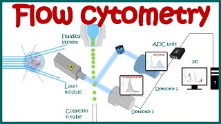
Is it that pc converts light intensity to voltage
Currently reviewing this for a job interview as a Flow Cytometry Technician
Great explanation..
Perfect, thanks
Nice presentation
Outstanding
At 4 minutes, does the data go directly to a PC, or is there a logic controller involved?
Very useful
Pls pls upload a video on circular dichromism
Thank You!
Quick question for my general understanding? At around 03:00 you say that the greater the cell, the more light will fall on the detector and vice versa for smaller cells.
My question is; Shouldn't it be the other way round, since the size of the detector won't change and the light will scatter more in other directions, thus leaving a larger 'shadow' behind on the detector?
Would appreciate an answer and thus thank you in advance.
However, thanks for the effort and the very nice demonstration on how this works! Stay motivated and curious! =)
Scattering appears not from the "borders" of the cell as it is in the pictures here, but from different intristic parts of the cell. So, if you have a larger cell, your detector will get scattered signal for longer time, thus the width of the forward signal peak (which is normally refered as FSC-W) wiil be greater than it is for a smaller cell. The height of the forward scattering peak (FSC-H, the maximum amount of light scattered during an event) could not be directly associated with sizes of the cells, but could be a marker which is unique for every type of cells. Normally we want to take in account both parameters (size and "type"), so we use their combination which is an area under the peaks curves (FSC-A).
nice bro
So helpfull for me
So small molecules will give less FW scatter in comparison to large molecules? How does that work? I always thought it was the other way around; big molecules block more light then small molecules so give less FW scatter. Can u explain it to me pls?
The cells are so small to block the laser beam ... the small cells cause less forward scatter and the large cells cause more scatter (as shown in the video between 2:50 and 3:00) ...
It's less "block more light" and instead scatters the light. So as was said before, all of the cells are too small to block the laser beam entirely. But, instead, as they pass through, they change the angle of the light so that it's not going straight, but scattering it. But think of it like a water tap. If you pass a small thing through it (your finger, for example), the flow breaks and spreads out. But if you pass something much larger (your fist, for example), the same thing happens, but to a much larger area. So, a small cell will scatter/spread out the light as it passes through it, but a bigger cell makes a much larger area of light scatter as it passes through. Hopefully that made sense...
So basically forward scattering depend upon size while side scattering depend on density.
Forward scatter depends on size and side scatter depends on granularity, or cell complexity, but not density.
Thanks so much
Thank you 🙏🙏
Thanks alot.
thankyou so much
how can chromosomes be sorted by flow cytometry..?
Thank yoooou ! 😁
Thank you
Thanks 👍
Thanks sis
Very well explained thank!
who i can detect the number of WBCs
Thank u
More video immunofluorescence,emsa,immunodiffusion,immunochemistry,
so far so good ,but where are the erythrocytes?
Hei, you can extract leucocytes from a blood sample in the laboratory ..
Usually the red blood cells are lysed and removed before running cells through the flow cytometer. In addition, we often set the threshold of a flow cytometer too high so that RBCs are excluded. If you were to keep the erythrocytes in the sample and set your threshold low enough to see them, they are very small and not granular, so they would show up at the very bottom left corner.
you need to revise it properly
You have amazing clarity with concepts but you need to work on delivery part, it gets to the point where it pisses off humongously
umm which i have aaaaa an example of i i i have aaaa an aaaaaa example of red blood cells........
I think most important is that she explains everything very clearly and it's easy to understand and remember. There is a lot youtubers with better oral skills but their explanations isn't so easy to follow. So I prefer her "aaaaa" that some else's fluency
Such aberrated speech...can't focus at all...myself, being a stammerer, I re-record the broken parts of voice over as many times as required...and that is important!
Your oration is very poor. Try to improve
You didn't do the best at explaining, a lot of "uhm"s and "uhhh"s, you were lost for your words half the time. In saying that the graphics is what saved this. I came here for a quick review so I knew what you were trying to say. I'd prepare a script for your next video.
Excellent explanation! Thank you
Thank you
Thank you