Corticospinal tracts/ Pyramidal Tracts Pathway |Neurophysiology| Super Easy Explanation

HTML-код
- Опубликовано: 2 сен 2020
- The descending tracts are the pathways by which motor signals are sent from the brain to lower motor neurones. The lower motor neurones then directly innervate muscles to produce movement.
The motor tracts can be functionally divided into two major groups:
Pyramidal tracts - These tracts originate in the cerebral cortex, carrying motor fibres to the spinal cord and brain stem. They are responsible for the voluntary control of the musculature of the body and face.
Extrapyramidal tracts - These tracts originate in the brain stem, carrying motor fibres to the spinal cord. They are responsible for the involuntary and automatic control of all musculature, such as muscle tone, balance, posture and locomotion
There are no synapses within the descending pathways. At the termination of the descending tracts, the neurones synapse with a lower motor neurone. Thus, all the neurones within the descending motor system are classed as upper motor neurones. Their cell bodies are found in the cerebral cortex or the brain stem, with their axons remaining within the CNS.
\
Pyramidal Tracts
By TeachMeSeries Ltd (2020)
Fig 2 - The medullary pyramids
The pyramidal tracts derive their name from the medullary pyramids of the medulla oblongata, which they pass through.
These pathways are responsible for the voluntary control of the musculature of the body and face.
Functionally, these tracts can be subdivided into two:
Corticospinal tracts - supplies the musculature of the body.
Corticobulbar tracts - supplies the musculature of the head and neck.
We shall now discuss both pathways in further detail.
Corticospinal Tracts
The corticospinal tracts begin in the cerebral cortex, from which they receive a range of inputs:
Primary motor cortex
Premotor cortex
Supplementary motor area
They also receive nerve fibres from the somatosensory area, which play a role in regulating the activity of the ascending tracts.
After originating from the cortex, the neurones converge, and descend through the internal capsule (a white matter pathway, located between the thalamus and the basal ganglia). This is clinically important, as the internal capsule is particularly susceptible to compression from haemorrhagic bleeds, known as a ‘capsular stroke‘. Such an event could cause a lesion of the descending tracts.
After the internal capsule, the neurones pass through the crus cerebri of the midbrain, the pons and into the medulla.
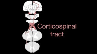
In the most inferior (caudal) part of the medulla, the tract divides into two:
The fibres within the lateral corticospinal tract decussate (cross over to the other side of the CNS). They then descend into the spinal cord, terminating in the ventral horn (at all segmental levels). From the ventral horn, the lower motor neurones go on to supply the muscles of the body.
The anterior corticospinal tract remains ipsilateral, descending into the spinal cord. They then decussate and terminate in the ventral horn of the cervical and upper thoracic segmental levels.
The corticobulbar tracts arise from the lateral aspect of the primary motor cortex. They receive the same inputs as the corticospinal tracts. The fibres converge and pass through the internal capsule to the brainstem.
The neurones terminate on the motor nuclei of the cranial nerves. Here, they synapse with lower motor neurones, which carry the motor signals to the muscles of the face and neck.
Clinically, it is important to understand the organisation of the corticobulbar fibres. Many of these fibres innervate the motor neurones bilaterally. For example, fibres from the left primary motor cortex act as upper motor neurones for the right and left trochlear nerves. There are a few exceptions to this rule:
Upper motor neurones for the facial nerve (CN VII) have a contralateral innervation. This only affects the muscles in the lower quadrant of the face - below the eyes. (The reasons for this are beyond the scope of this article)
Upper motor neurons for the hypoglossal (CN XII) nerve only provide contralateral innervation.


Thank you so much ❤
Very easy to understand. Thank you so much 🙏❤️
Tq mam ur video helped me during revison time of my exams😊😊
This video was very helpful thank you 😊
You're welcome 😊
Thank you u made it easy .Please can you make video on cortico-neuclear tract and cortico-muscular tract?
Thanks ma’am ☘️☘️☘️
Nice presentation 👏 thankyou ❤
Thank you soo much.🙏🙏 Crystal clear 😊pls upload more ma'am 😊
You're welcome 😊, sure
simply awesome explanation ❤
Very very good mam ❣️❣️❣️
This was so easy to understand! Thank you
You're welcome 😊
Crystal clear explanation mam....plz upload more videos on CNS physiology mam
Thank you 😊, will be uploading shortly
thankyouu❤
Thanks 😊👍
Thank you sister!! It is very helpful to me🙂
Amazing and very useful video 🤩
Thank you
You're welcome 😊
Excellent Presentation !! DR RIZVI NEURO-S
It was really helpful thank you🙏🙏🌸
You're welcome 😊
Awesome
Good teaching mam
Thank you, keep watching ❤️
Well explained 🌸
Thank you 😊
Thank you so much ❣️
Don't know why I was fearing from this topic
Thank you
Which book you referred?
Sembu
Helpful ❤️
Automatic shivering can also be told as tremers...
excellent madam. unknowingly you said instead of saying cerebrovascular accident said cardiovascular accident
Tq plz upload extrapyramidal tracts also
Can you explain basal ganglia and pain pathway
Please make video on Ascending tracts also..🙏👍
I have 1 question...Corticospinal tract k 90 % fibre decussate kar jate h medulla se
To aisa hoga hoga kya..ki decussation se pehle lesion hoga to contralateral symptom ayenge or decussation k bad lesion hoga to ipsilateral symptom ayenge??
Thanku mam❤️
You're welcome 😊
Gud mam 👌 👍 😊 😀
Video has nice content and made easy to understand........but one thing the background music is disturbing a loooootttt plz fix it
Gamma motor neurons at termination
This is from sembulingam
Haa
Line to line from sembulingam
Awesome
Thank you ❤️