3D Middle Ear & External Ear embryology - Impedance matching - Ossicles Embryology

HTML-код
- Опубликовано: 27 июл 2024
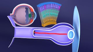
- In this visual medical lecture Dr. Aizaz from MedicoVisual talks about the development of tympanic cavity (middle ear cavity), pharyngotympanic tube, epitympanic recess, middle ear ossicles, external ear (external auditory meaturs and pinna or auricle) and the physiology of impedance matching mechanism.
00:00 Introduction
00:43 Why is there a need of middle ear cavity and external auditory meatus
04:37 Development of External Auditory Meatus from Dorsal end of First Pharyngeal cleft
07:40 Tubotympanic recess and development of middle ear cavity and auditory tube
12:09 Impedance Matching mechanism (basic physiology)
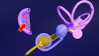
13:51 Development of Middle Ear Bones (Ossicles; Malleus, Incus and Stapes)
19:44 Embryology of epitympanic recess and middle ear ligaments
22:33 Mastoid Air cells and Mastoid Antrum
23:50 Stapedius and Tensor Tympani muscle
28:53 Tympanic Membrane Embryology
30:19 Pinna / Auricle of the external ear


👂📚 This video is a part of our "Premium 3D Ear Embryology" course on MedicoVisual! Dive into the complete course for FREE on our website: www.medicovisual.com/courses/premium-3d-ear-embryology
Don't miss out on in-depth, 3D-animated lectures and a comprehensive understanding of ear development. Join us in this educational journey! 🌟
🔗 Click the link above to access the full course. Happy learning! 🧠📖
You are really no one but a god for the med students as embryology is a subject to be visualised in 3d for proper understanding and somehow the textbooks and normal college lectures have consistently failed to make the 3d concept and making it further hard to understand the paraclinical and clinical subjects.... thanks a lot Sir/Saviour.
I am honored 🤗
I wanted to express my sincere gratitude for your lectures. Your passion for the subject matter shines through, making the material engaging and enjoyable to learn. Your clear explanations and dedication to helping students truly enhance the learning experience. Thank you for being such an inspiring educator.
Wow thank you so much for such inspiring words. Made my day 😊😊😊
Sir ,in my college they didn't teach embryology much , just from faq the topics were taken , by your video only Iam getting to know all information 🙏thank you so much sir
Well done! Thank you for your tremendous efforts!
Thanks. It's very helpful and easy to memorize.❤❤❤❤
Well done
Thank you so much
subhan allah
Mind blowing ❤
Thank you so much
Thanks
❤
🔥
❤❤❤
❤❤❤❤
If you turn the stapes upside down like you did would it be solid or is it an opening in the middle of the stapes structure. When it's upside in the middle when you look down towards the head of the stapes is there an opening to the head or is it solid?????
I am sorry, I did not quite understand your question. Please elaborate.
How are you doing this graphics and all??? Which app is this??? Where did u learn these things??? U r such a genius bro!!
Thank you so much.
I use Blender (www.blender.org) for 3D animations and PowerPoint for 2D animations. I started learning these technologies during my student life because I love teaching and I had envisioned MedicoVisual even during my student life.
@@MedicoVisual very nice bro ❤️🔥❤️🔥 all the best
❤🙌
Upload animation video of anterior & posterior malleolar flod of tympanic membrane
Dear, Malleolar folds are topic of Anatomy. We will discuss that in anatomy of ear lectures later someday