Neck Fascia

Поделиться
HTML-код
  • Опубликовано: 16 сен 2024
  • This video briefly describe the arrangement of neck fascia that introduces to fascial spaces and spread of dental infection.
    1. Superficial Fascia: encloses platysma and muscles of facial expression
    2. Deep Fascia is arranged in 3 main layers:
    a. superficial investing layer that invests trapezius and sternoclidomastoid muscles as well as submandibular and parotid glands.
    b. Middle layer (Pretracheal Fascia): it has visceral part that encloses thyroid gland, trachea and esophagus and the muscular layer that encloses the infrahyoid muscles
    c. Deep layer that forms of alar and prevertebral layers.
    This arrangement of deep fascia creates several spaces that could be potential route for spread of dental infection through the neck to mediastinum.
    Important spaces"
    1. Sublingual
    2.Submandibular
    3. Masticatory space that divided into; massetric, ptergomandibular and temporal
    4. Parapharyngeal
    5. Retropharyngeal
    6. Danger
    7. Prevertebral
    8. Carotid vascular space
    9. Canine space
    10. buccal

Комментарии • 80

  • @dikshadinker4313
    @dikshadinker4313 3 месяца назад

    12 years of my medical education and I still come back to your videos!

  • @grouchmy8734
    @grouchmy8734 Год назад +1

    thank you for making my life easier

  • @hanyelhag1664
    @hanyelhag1664 4 года назад +4

    Absolutely wonderful and well explained .. can't thank you enough sir

  • @hh-zq9io
    @hh-zq9io 5 лет назад

    Cant thank u enough Sir....plz dont stop doing such videos.....million thx,,,u saved my exam

  • @haiderbokhari6151
    @haiderbokhari6151 5 лет назад +4

    brilliant... absolutely brilliant.

  • @02-adhiparnasaha81
    @02-adhiparnasaha81 5 лет назад +2

    Now my concepts are crystal clear. Thank you Sir 😁😁

  • @umerbinshabir6561
    @umerbinshabir6561 5 лет назад +2

    I wish you had more videos sir......thank you so much sir....

  • @lucillemeisner251
    @lucillemeisner251 3 года назад +2

    Thank you; very helpful!

  • @yazanahmad8926
    @yazanahmad8926 6 лет назад +1

    thank you very much doctor you are very helpful but i wish if you have more video i like your way in Explanation you are the best

  • @rubasarkez9917
    @rubasarkez9917 5 лет назад +2

    Amazing. You've made it so easy to me ❤❤❤❤ thanx a lot

  • @arezoomg8196
    @arezoomg8196 6 лет назад +1

    It’s perfect. completely explained.Thank you so much

  • @Synth009
    @Synth009 6 лет назад

    Tnk u a lot. New subscriber. You should get more viewers and followers, made my learning very easy

  • @thcfly94
    @thcfly94 7 лет назад +1

    great video helped me a lot revising, thank you

  • @mahmoudhelmy5287
    @mahmoudhelmy5287 3 года назад

    You are amazing!
    Thank you a lot Dr.

  • @gtan06
    @gtan06 6 лет назад +1

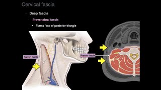
    Pre-tracheal layer attaches to hyoid bone superiorly as well as oblique line thyroid cartilage

  • @naremantarek6787
    @naremantarek6787 5 лет назад +1

    life saving-ly explained! thank you

  • @pratikparajuli6167
    @pratikparajuli6167 4 года назад

    superb

  • @aribaqamar8258
    @aribaqamar8258 3 года назад

    amazing!

  • @syedafarhatfatima4091
    @syedafarhatfatima4091 4 года назад

    Awesome, sir u really make this topic easy... Keep uploading sir.

  • @harithareddy2941
    @harithareddy2941 6 лет назад +1

    Great explanation sir 👍👍👍👍

  • @najihm.t7484
    @najihm.t7484 7 лет назад

    Really helpful.....very nice explanation.....thanku so much.......

  • @justinenyangaresi4174
    @justinenyangaresi4174 4 года назад

    Well explained. Keep up with the good work.

  • @justinenyangaresi4174
    @justinenyangaresi4174 7 лет назад

    Very good and helpful explanation

  • @isaacheron1607
    @isaacheron1607 5 лет назад

    Well arranged. Very helpful. Thank you sir.

  • @victorstark8857
    @victorstark8857 6 лет назад +2

    Great video tutorial
    Thank you sir for your effort!@Easy Anatomy

  • @aishanawzad3781
    @aishanawzad3781 6 лет назад +1

    Thank you very much
    Really great video ☘️☘️

  • @azizatarmissi1907
    @azizatarmissi1907 3 года назад

    Amazing

  • @DrAnkit159
    @DrAnkit159 7 лет назад +1

    thanks a lot ...video helped me growing concept

  • @maryamesmaeili7592
    @maryamesmaeili7592 7 лет назад +2

    Hence da name, easy anatomy!
    Thank you indeed,

  • @ara_wy
    @ara_wy 7 лет назад +1

    it's very helpful. thanks a lot!

  • @Dr_Fantacyfactory_
    @Dr_Fantacyfactory_ 5 лет назад +2

    GRT explanation sir plz upload vedios on infratemporal fossa

    • @EasyAnatomy
      @EasyAnatomy  5 лет назад

      Thank you!. You can learn the detail of infratemporal fossa If you watch the videos of skull, muscles of mastication, mandibular nerve, maxillary artery and otic ganglion. All these together represent infratemporal fossa.
      Best luck!

  • @nikitashah3224
    @nikitashah3224 6 лет назад

    Thank uu so much
    Explanation is very very good👍👍👍

  • @ankushjain4986
    @ankushjain4986 5 лет назад +1

    Thanks a lot !! Really helped

  • @Allo_veda
    @Allo_veda 6 лет назад

    Very helpful video
    Thanks a lot

  • @maurelletallak469
    @maurelletallak469 3 года назад

    It was helpful.thank u

  • @marinakamal5812
    @marinakamal5812 3 года назад

    Amazing one .. many thanks 🌼🌼

  • @hamzehzghool6152
    @hamzehzghool6152 7 лет назад +1

    Great ..you're great sir !

  • @KunalM-ec3pm
    @KunalM-ec3pm 5 лет назад

    Thank you so much! Very well explained

  • @shamakuma1967
    @shamakuma1967 5 лет назад

    Also facial nerve branches like temporal, transverse cervical, zygomatic, buccal, mandibular.

  • @karandeepgill186
    @karandeepgill186 6 лет назад

    Awesome video

  • @akshittuli1211
    @akshittuli1211 7 лет назад +1

    thanks, nice explanation !!

  • @saiedrahman5120
    @saiedrahman5120 7 лет назад

    Great video thank you so much

  • @chantzenian
    @chantzenian 6 лет назад

    Great video!

  • @hina5243able
    @hina5243able 3 года назад

    why does not a tumor spread up to the hyoid bone, when the pretracheal fascia goes upto the hyoid bone? it has an attachment in the middle line to the hyoid bone, although the oblique line stops at the thyroid cartilage? (would appreciate an answer , hope my question is understandable!!)

  • @mustafamukaram7412
    @mustafamukaram7412 6 лет назад +1

    Ma sha allah 😍😍😍

  • @mawaddashaban477
    @mawaddashaban477 6 лет назад +1

    Thank u so much ,
    Just I want to mention our doctor said that Pretracheal Fascia attaches superiorly to hyoid bone .

    • @EasyAnatomy
      @EasyAnatomy  6 лет назад +2

      Yes, he is correct it attaches to both thryroid cartilage and also to hyoid bone

  • @umair3899
    @umair3899 5 лет назад

    Thanks alot sir... You are a saviour ❤❤❤

  • @ossamazakaria4760
    @ossamazakaria4760 5 лет назад

    thank you very much great explanation

  • @husseinimad1354
    @husseinimad1354 4 года назад

    Good

  • @amishakanwar7942
    @amishakanwar7942 6 лет назад +1

    Good video thanks

  • @mohamedelshamaa2621
    @mohamedelshamaa2621 5 лет назад

    Perfect

  • @dr_ismail_wazir
    @dr_ismail_wazir 4 года назад

    much appreciTed , ty

  • @peeyushrawat6052
    @peeyushrawat6052 4 года назад

    Thank you sir

  • @hh-zq9io
    @hh-zq9io 5 лет назад

    Million thx....u r a saver

  • @SohelRana-ox5rc
    @SohelRana-ox5rc 5 лет назад

    Nice to see

  • @dikshadinker4313
    @dikshadinker4313 3 года назад

    💯

  • @drpallaveeghabak8129
    @drpallaveeghabak8129 5 лет назад

    thank u so much 😊

  • @kosharicorner157
    @kosharicorner157 5 лет назад

    Thanks alot

  • @missaster1902
    @missaster1902 5 лет назад

    Nice mash aAllah

  • @rohan1632
    @rohan1632 4 года назад

    Thanks sir

  • @MarcosJ-mq4lk
    @MarcosJ-mq4lk 7 лет назад

    I have fascia damage/worn on right occipital area with horrendous,at times,pain!/./.
    Ive been revered to a neurologist....what would they do/?

  • @zahraamahmoud5661
    @zahraamahmoud5661 6 лет назад

    Thanks

  • @umerbinshabir6561
    @umerbinshabir6561 5 лет назад +2

    I wish I could write it in the same way in exam😍😍😍😍

  • @bushrawadani3420
    @bushrawadani3420 5 лет назад

    Thnks alot

  • @Swaad172
    @Swaad172 7 лет назад

    vry much helpful

  • @lolynice71
    @lolynice71 6 лет назад

    thank u sooooo much

  • @doctorshocker619
    @doctorshocker619 6 лет назад

    You are BOMB 👌👌👍👍👌👍

  • @likarao5864
    @likarao5864 6 лет назад

    tnx sir

  • @MustafaKhan-sr3rr
    @MustafaKhan-sr3rr 7 лет назад

    U havent explained axillary sheath

  • @ayub.m.shalan7246
    @ayub.m.shalan7246 6 лет назад +1

    good

  • @anghammarea4335
    @anghammarea4335 7 лет назад

    رائع😍😍😍😍

    • @EasyAnatomy
      @EasyAnatomy  7 лет назад

      Thank you! glad to know it helps

  • @ahmedhatem489
    @ahmedhatem489 6 лет назад

    ارجوك ماتكتبش بالاقلام دي تاني عشان بتقشعرني وبتأذيني فشخ

  • @ayushraj5782
    @ayushraj5782 5 лет назад

    The idea of writing everything with sketch pen is bad.

    • @EasyAnatomy
      @EasyAnatomy  5 лет назад

      Any suggestion? thank you!

    • @ayushraj5782
      @ayushraj5782 5 лет назад

      You explained well, but many peope get irritated by the sound of sketch pen rubbing on paper, though everyone doesnt have this problem. I suggest you to use ball/ink/gel pen along with color pencils. Thank you.