This is masterpiece. Dr. Anagha Joshi madam is my mentor and guide. Many students were always envious of not getting to train under Madam. This will give them the peek of how fine and elaborative the teaching sessions Madam takes. People are lucky for the digital platform to reach this precious material globally.
Thanks madam for this excellent presentation, one of the best I have watched in recent time. Your students are really blessed to have a teacher like you. 🙏🙏
Today I did my 1st 2 temporal bone CT scans and both of them were finalised with minimal changes by professors.Thank u soo much for this video..Had been watching this for the past 1 day and trying to learn HRCT temporal bone.Thank u soo much ma'am
Madem i have deviated nasal septum on left side of nose. And at the same time i have tinitus to my Righr ear ( 24 hrs ringing like sea shore). From last 25 years. MRI BRAIN NORMAL. WHICH PART OF CT OR MRI ( POSITION) I TAKE TO EVALUATE TINITUS OF MY RIGHT EAR?
Very well presented mam.. can’t get substitute must say the excellent one ..... mam can I get to know lectures on laryngeal anatomy and it’s pathologies plus head n neck anatomy and it’s pathology too presented by you...
Mam in Head CT Scan Report it is only written--'' Fracture Right Temporal Bone '' then how it can be known that out of 5 parts of temporal bone which part is fractured.
Respected Mam...i have 2 questions regarding hrct of temporal bone 1. Are temporal bone hrct is always done for both sides same time and 2. Is the radiation dose is same or different in left temoral bone or right temporal bone...?


This is masterpiece. Dr. Anagha Joshi madam is my mentor and guide. Many students were always envious of not getting to train under Madam. This will give them the peek of how fine and elaborative the teaching sessions Madam takes. People are lucky for the digital platform to reach this precious material globally.
Thanks for your comment Atish!
best video for a beginner. absolutely marvelous.
Glad you liked it
One of the best anatomy illustration . Thank u so much mam🤗
I’m an ENT surgeon maam. But always love listening to your lecture. Every time I listen to it I feel I learn something new.
Best anatomy lecture on temporal bone.
Must see video for radiology residents
Thank you for this mam , no one teaches temporal bone like you !
Thanks for liking Nakul
Great video, very helpful... Thank you.
All time favourite madam 👍👍
Very nice informative..
Thanks a lot very informative Lecture )) Greetings from Germany ))
A fantastic presentation ! Thank you.
Really really thank you so much maam....no one teaches like you.
So nice and informative lecture mam.
I am lucky being your Student.
Thanks mam.
Great lecture ma'am..very informative and crisp..thank you.
well said..everything in nutshell..love itt....
Thanks madam for this excellent presentation, one of the best I have watched in recent time. Your students are really blessed to have a teacher like you. 🙏🙏
Clear and beautiful description of anatomy..appreciations ftom an ENT surgeon, doctor!!
Most welcome!
Today I did my 1st 2 temporal bone CT scans and both of them were finalised with minimal changes by professors.Thank u soo much for this video..Had been watching this for the past 1 day and trying to learn HRCT temporal bone.Thank u soo much ma'am
Amazing explanation Mam. ❤ Middle ear anatomy has always been difficult. You made it very easy. Chakli 😂 you are definitely from Karnataka mam
Very nicely explained …thank you !!!
Very very informative lecture.Thank you madam
thanks very much, so informative lecture 👍
Excellent
Amazing
And
Awesome Ma’am
Congratulations 👏🎉 😊
Awesome, just awesome
thanks Radness, we like your name 😍😍
Very Impressive👌👌
Excilently explained in detail,
Great lecture
Amazing video maam✌️
Loved it.
nice informative video.
Very nice and informative lecture....
Thanks a lot Ashok
Excellent
Awesome Lecture Ma'am
Thanks for liking
Thank you
Brilliant madam 😄
superb mam ...
Thank you so much madam.
You are most welcomeAmit
Very knowledgeable video .. thanks
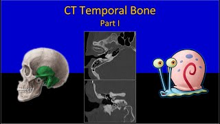
HRCT of the temporal bone with thin sections (1l.0 mm) along with plain coronal & axial
images were ta
Thank u all
Madem i have deviated nasal septum on left side of nose. And at the same time i have tinitus to my Righr ear ( 24 hrs ringing like sea shore). From last 25 years. MRI BRAIN NORMAL. WHICH PART OF CT OR MRI ( POSITION) I TAKE TO EVALUATE TINITUS OF MY RIGHT EAR?
Same problem, how are u now, plz reply
thanks a lot
Thanks Ma'am
Very well presented mam.. can’t get substitute must say the excellent one ..... mam can I get to know lectures on laryngeal anatomy and it’s pathologies plus head n neck anatomy and it’s pathology too presented by you...
Mam in Head CT Scan Report it is only written--'' Fracture Right Temporal Bone '' then how it can be known that out of 5 parts of temporal bone which part is fractured.
Madan temporal bone brain mri me dikhta h?
will this be saved for watching it again later?
Yes
yes, all videos are available for viewing after the premiere, and downloadable on your device if yo9u need to. Thanks for watching
Thank you
Dr ñ k bhattacharjee
Narashinghapur
Respected Mam...i have 2 questions regarding hrct of temporal bone 1. Are temporal bone hrct is always done for both sides same time and 2. Is the radiation dose is same or different in left temoral bone or right temporal bone...?
Same time
So one time radiation
@@anaghajoshi3966 thanks mam for the information
Kindly apna pointer to visible rkhen
Itna chota hy k nazr hi ni ata