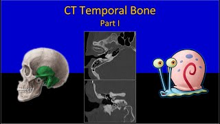
HRCT TEMPORAL BONE- Illustrations and detailed radioanatomy on HRCT. FACIAL NERVE on HRCT T BONE.

HTML-код
- Опубликовано: 10 июл 2024
- We discuss anatomy of external, middle and inner ear including facial nerve with illustrations and neat labelled diagrams. Detailed radiological anatomy of Middle ear ossicles.
We scroll through axial and coronal HRCT temporal bone section by sections and locate all structures with detailed imaging anatomical review.
Check out our other radio-anatomy videos
Larynx - • LARYNX ANATOMY - Illus...
Liver- • LIVER SEGMENTS RADIOAN...
Cerebral venous anatomy- • CEREBRAL VENOUS ANATOM...
Cerebral arterial anatomy - • ARTERIAL ANATOMY OF BR...
Pituitary gland - • RADIO-ANATOMY OF PITUT...


Best video with illustrated diagrams for understanding 😊
Thanks a lot 🙏
Classical explanation,
Thank you for this well informative video ❤