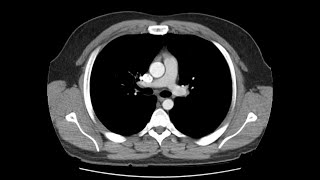
Anatomy of a Transverse CT of the Thorax

HTML-код
- Опубликовано: 6 фев 2025
- An overview of the anatomy visible in a transverse computed axial tomographical image of the thorax (and part of the abdomen) performed with intravenous contrast and acquired in the arterial phase.
[The CT image was uploaded to Radiopedia by Dr Andrew Dixon
radiopaedia.or...]


This is the best thorax axial CT lecture on RUclips. You gave great amount of detail without using complex words. Illustration of structures with colors makes it very easy to learn. Thank you very much for this video.
Best CT video I've ever found on RUclips. Thank you very much, you're a hero.
Amazing video. You should feel proud that 8 years later your work keeps helping students around the world! Thank you sincerely from Argentina ♥
Thank you for your work, I'm a Brazilian student currently in the second semester, and this is really helpful!
I have been looking for someone to show the axial ct of thorax at vertebral levels for hours and finally found yours
Thank you so much Dr. Eva... I just wasn't understanding from what I was reading in my book... this video helped clear up a lot of questions I was having. Much appreciated. -Rad tech student.
Cant thank you enough. What a concise and yet so informative.. hit the nail. Appreciate your hardwork. Thanks alot
Thank you 1000.....It is so helpful,so accurate,so brilliant,so easy approach, so much correlatable, so much clear cut margins, so much explanation, so much perfection.....and yes the voice was 🔥 fire......Thank u so so much....Thank God,I found it after struggling for 1long year....😢
Best explanation on thorax axial CT, thank you
Thankyou Dr.Eva Sweeney, . Its a best way to learn these Thoracic CT anatomy. ( From Nepal)
Thank you Dr Eva for explaining in such a good way...
Waiting for your next Lectures on different parts of the body....
A freshly technologist from Pakistan
Woooow never thought CT chest can be so easy, thank you. Great work ❤❤❤❤❤
We miss you and your Videos so much.. Grweat teacher
Thank you Dr. Eva.. wonderful presentation
Thank you Dr. Eva Sweeney. Very detailed and helpful education material.
This was a fantastic video! Extremely helpful. Thank you!
Perfectly labelled radiologic anatomy :)
best CT Thorax explenatiion on youtube 😍
wow it's so nice and helpful to understand the important information in a easy way.....thank you doc😊
Much better than many of the good radilogy video.Marking each organ make understading much easy.Not sure if you can do similar rad video with markings on common pathology.
This is super clear and helpful! I will be waiting for your next video on the abdominal ct 🙂
Thanks for providing us the link for online Radiology Courses...
Nice video, I liked the editing especially
This video was so helpful, thank you very much!
I hope more video’s like this for the other parts specially fot the head 🥹💕 and thank you so much u helped me a lot i’m from Ksa 🇸🇦
Thank you Dr eva great work
Vey concise and informative ❤
This video is like a dream come true
Please please make a similar one on CT abdomen. Thank you
This was great. Thank you doc
I am indian to following your teaching online class 🙏
Thank you very much Dr.Eva
Thanks for the very good elaboration 👍
Incredible work! Really helpful! Thank you!
Excellent explanation, thanks a lot !!
Just subscribed and loved ❤❤❤❤❤
Thanks for this work.
Very Helpful, Thank u very much
Thanks !
Can u plz upload a video for anatomy of abdominal ct scan
Awesome video
Thanks
Thank you so much...this is sooooo clear and detailed
Thanks doc
Amazing work! This will be helpful for lots!!
Great job 👏
really that is one of the great illustrating and educational video. Can you make more like this one? especially for the head and neck
Excellent presentation ! Thank you so much.
this was just perfect! thank you
love you for this wonderful presentation.
Very well taught video.Loved it.Thank you.
Simple and lucid 😊
Good work.....❤
I liked that u highlighted each part in different colors. I hate that you started inferiorly and worked your way up. It makes it harder for me to follow along since every other tutorial starts top then goes to bottom.
Dr Eva thanks enough
Awesome would please talk about the CT of abdomen and other organs
Thanks a million!
Very helpful
Excellent and good for education purpose. I learned a lot. I need segital n lung window
It is so helpful thank you
Thank you!
Great video, thank you.
Brilliant and very useful 💕
you are awesome evinha
amazing, thank u very much!
Colon or large intestine ulcers can be seen in ct scan?
Thank you miss
Syed umer Farooq from kashmir
Very helpful for me thanks
Good teaching 🙏
Thanks doc.
Thank you
Good teaching mem 👍
incredible, thank you !
Thank you.
Thank you so much, very helpful
thx, very useful
Very good
thank you so much
Very educational!!!!!!
Wonderful, thank you
Awesome 👏🏻 thanks
Please discuss H rct of cheast.
You are an angel 😇. Thanks.
Thanks 👍
Very nice.....informative
Very helpful.thanks
Not me watching this the day before my exam (': thank you
Thank you👍🏼
Thank u so much
Thank you ♥️
this was awesome thankyou
thx dr eva .
Excellent video Dr Eva.. Can a similar one be uploaded for CT anatomy of Neck.. Thnx
Thanks 😘😘
Great
My findings..thats why i cannot working abroad.pls help me.
Impression
1.vague opacities in bilateral upper lobes.biapical pleuroparenchymal fibrosis.these may be due to infection such a ptb.followup complete resolution
2. Tiny centrilobular nodules in tree in bud apperance in bilateral upper lobes may be due to bronchiolitis,or endobronchial spread of disease.focal air trapping in periphery of left upper lobe
hey dr.eva sweeney. ıts wonderfull video. can you tell us on abdomen ct ? thank you
Great 👍
Question, can the lung parenchyma not been seen on this scan?
Thank youuu
Really helpful
Dr eva can I send you a short video of a ct so that you could explain to me what is that I see and is not on your video. Thanks